• 文献检索
  • 文档翻译
  • 深度研究
  • 学术资讯
  • Suppr Zotero 插件Zotero 插件
  • 邀请有礼
  • 套餐&价格
  • 历史记录
应用&插件
Suppr Zotero 插件Zotero 插件浏览器插件Mac 客户端Windows 客户端微信小程序
定价
高级版会员购买积分包购买API积分包
服务
文献检索文档翻译深度研究API 文档MCP 服务
关于我们
关于 Suppr公司介绍联系我们用户协议隐私条款
关注我们

Suppr 超能文献

核心技术专利:CN118964589B侵权必究
粤ICP备2023148730 号-1Suppr @ 2026

文献检索

告别复杂PubMed语法,用中文像聊天一样搜索,搜遍4000万医学文献。AI智能推荐,让科研检索更轻松。

立即免费搜索

文件翻译

保留排版,准确专业,支持PDF/Word/PPT等文件格式,支持 12+语言互译。

免费翻译文档

深度研究

AI帮你快速写综述,25分钟生成高质量综述,智能提取关键信息,辅助科研写作。

立即免费体验

基于人工智能算法的宫颈癌多模态 MRI 分析。

Multimodal MRI Analysis of Cervical Cancer on the Basis of Artificial Intelligence Algorithm.

机构信息

Department of Obstetrics and Gynecology, Xi'an Daxing Hospital, Xi'an 710000, Shaanxi, China.

Department of Obstetrics and Gynecology, Affiliated Hospital of Yan'an University, Yan'an 716000, Shaanxi, China.

出版信息

Contrast Media Mol Imaging. 2021 Nov 8;2021:1673490. doi: 10.1155/2021/1673490. eCollection 2021.

DOI:10.1155/2021/1673490
PMID:34858113
原文链接:https://pmc.ncbi.nlm.nih.gov/articles/PMC8592750/
Abstract

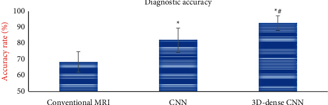
The purpose of this study is to explore the application value of artificial intelligence algorithm in multimodal MRI image diagnosis of cervical cancer. Based on the traditional convolutional neural network (CNN), an artificial intelligence 3D-CNN algorithm is designed according to the characteristics of cervical cancer. 70 patients with cervical cancer were selected as the experimental group, and 10 healthy people were selected as the reference group. The 3D-CNN algorithm was applied to the diagnosis of clinical cervical cancer multimodal MRI images. The value of the algorithm was comprehensively evaluated by the image quality and diagnostic accuracy. The results showed that compared with the traditional CNN algorithm, the convergence rate of the loss curve of the artificial intelligence 3D-CNN algorithm was accelerated, and the segmentation accuracy of whole-area tumors (WT), core tumor areas (CT), and enhanced tumor areas (ET) was significantly improved. In addition, the clarity of the multimodal MRI image and the recognition performance of the lesion were significantly improved. Under the artificial intelligence 3D-CNN algorithm, the Dice values of WT, ET, and CT regions were 0.78, 0.71, and 0.64, respectively. The sensitivity values were 0.92, 0.91, and 0.88, respectively. The specificity values were 0.93, 0.92, and 0.9 l, respectively. The Hausdorff (Haus) distances were 0.93, 0.92, and 0.90, respectively. The data of various indicators were significantly better than those of the traditional CNN algorithm (  0.05). In addition, the diagnostic accuracy of the artificial intelligence 3D-CNN algorithm was 93.11 ± 4.65%, which was also significantly higher than that of the traditional CNN algorithm (82.45 ± 7.54%) (  0.05). In summary, the recognition and segmentation ability of multimodal MRI images based on artificial intelligence 3D-CNN algorithm for cervical cancer lesions were significantly improved, which can significantly enhance the clinical diagnosis rate of cervical cancer.

摘要

本研究旨在探讨人工智能算法在宫颈癌多模态 MRI 图像诊断中的应用价值。基于传统卷积神经网络(CNN),根据宫颈癌的特点设计了一种人工智能 3D-CNN 算法。选取 70 例宫颈癌患者作为实验组,选取 10 例健康人作为参考组。将 3D-CNN 算法应用于临床宫颈癌多模态 MRI 图像的诊断中,综合评价算法的图像质量和诊断准确率。结果表明,与传统 CNN 算法相比,人工智能 3D-CNN 算法的损失曲线收敛速度加快,全肿瘤区(WT)、核心肿瘤区(CT)和增强肿瘤区(ET)的分割准确率显著提高。此外,多模态 MRI 图像的清晰度和病变的识别性能也显著提高。在人工智能 3D-CNN 算法下,WT、ET 和 CT 区域的 Dice 值分别为 0.78、0.71 和 0.64,灵敏度值分别为 0.92、0.91 和 0.88,特异度值分别为 0.93、0.92 和 0.91,Hausdorff(Haus)距离分别为 0.93、0.92 和 0.90,各指标数据均明显优于传统 CNN 算法(  0.05)。此外,人工智能 3D-CNN 算法的诊断准确率为 93.11 ± 4.65%,也明显高于传统 CNN 算法(82.45 ± 7.54%)(  0.05)。综上所述,基于人工智能 3D-CNN 算法的宫颈癌病变多模态 MRI 图像的识别和分割能力显著提高,可显著提高宫颈癌的临床诊断率。

https://cdn.ncbi.nlm.nih.gov/pmc/blobs/5bfe/8592750/7fe3eb67bc04/CMMI2021-1673490.011.jpg
https://cdn.ncbi.nlm.nih.gov/pmc/blobs/5bfe/8592750/67e58bf2d57e/CMMI2021-1673490.001.jpg
https://cdn.ncbi.nlm.nih.gov/pmc/blobs/5bfe/8592750/9680bbcfa60a/CMMI2021-1673490.002.jpg
https://cdn.ncbi.nlm.nih.gov/pmc/blobs/5bfe/8592750/2c01e2ac31a6/CMMI2021-1673490.003.jpg
https://cdn.ncbi.nlm.nih.gov/pmc/blobs/5bfe/8592750/fb3a3886815f/CMMI2021-1673490.004.jpg
https://cdn.ncbi.nlm.nih.gov/pmc/blobs/5bfe/8592750/a5fc7ae7be37/CMMI2021-1673490.005.jpg
https://cdn.ncbi.nlm.nih.gov/pmc/blobs/5bfe/8592750/68c5a3291557/CMMI2021-1673490.006.jpg
https://cdn.ncbi.nlm.nih.gov/pmc/blobs/5bfe/8592750/2a4da034b9bf/CMMI2021-1673490.007.jpg
https://cdn.ncbi.nlm.nih.gov/pmc/blobs/5bfe/8592750/0f21e5e810d2/CMMI2021-1673490.008.jpg
https://cdn.ncbi.nlm.nih.gov/pmc/blobs/5bfe/8592750/47513df5baf0/CMMI2021-1673490.009.jpg
https://cdn.ncbi.nlm.nih.gov/pmc/blobs/5bfe/8592750/c79b05fa614d/CMMI2021-1673490.010.jpg
https://cdn.ncbi.nlm.nih.gov/pmc/blobs/5bfe/8592750/7fe3eb67bc04/CMMI2021-1673490.011.jpg

相似文献

1
Multimodal MRI Analysis of Cervical Cancer on the Basis of Artificial Intelligence Algorithm.基于人工智能算法的宫颈癌多模态 MRI 分析。
Contrast Media Mol Imaging. 2021 Nov 8;2021:1673490. doi: 10.1155/2021/1673490. eCollection 2021.
2
Diagnosis of Early Cervical Cancer with a Multimodal Magnetic Resonance Image under the Artificial Intelligence Algorithm.基于人工智能算法的多模态磁共振成像在早期宫颈癌诊断中的应用。
Contrast Media Mol Imaging. 2022 Mar 23;2022:6495309. doi: 10.1155/2022/6495309. eCollection 2022.
3
The Value of Convolutional Neural Network-Based Magnetic Resonance Imaging Image Segmentation Algorithm to Guide Targeted Controlled Release of Doxorubicin Nanopreparation.卷积神经网络磁共振成像图像分割算法指导阿霉素纳米制剂靶向控释的价值。
Contrast Media Mol Imaging. 2021 Jul 26;2021:9032017. doi: 10.1155/2021/9032017. eCollection 2021.
4
Deep Learning Algorithm-Based MRI Image in the Diagnosis of Diabetic Macular Edema.基于深度学习算法的 MRI 图像在糖尿病性黄斑水肿诊断中的应用。
Contrast Media Mol Imaging. 2022 Mar 4;2022:1035619. doi: 10.1155/2022/1035619. eCollection 2022.
5
Utilization of Ultrasonic Image Characteristics Combined with Endoscopic Detection on the Basis of Artificial Intelligence Algorithm in Diagnosis of Early Upper Gastrointestinal Cancer.基于人工智能算法的超声图像特征与内镜联合检测在早期上消化道癌诊断中的应用。
J Healthc Eng. 2021 Nov 29;2021:2773022. doi: 10.1155/2021/2773022. eCollection 2021.
6
Artificial Intelligence Algorithm-Based Computerized Tomography Image Features Combined with Serum Tumor Markers for Diagnosis of Pancreatic Cancer.基于人工智能算法的计算机断层扫描图像特征与血清肿瘤标志物联合用于胰腺癌的诊断。
Comput Math Methods Med. 2022 Mar 2;2022:8979404. doi: 10.1155/2022/8979404. eCollection 2022.
7
Colposcopic multimodal fusion for the classification of cervical lesions.阴道镜下多模态融合在宫颈病变分类中的应用。
Phys Med Biol. 2022 Jun 22;67(13). doi: 10.1088/1361-6560/ac73d4.
8
[Application of convolutional neural network to risk evaluation of positive circumferential resection margin of rectal cancer by magnetic resonance imaging].卷积神经网络在直肠癌磁共振成像环周切缘阳性风险评估中的应用
Zhonghua Wei Chang Wai Ke Za Zhi. 2020 Jun 25;23(6):572-577. doi: 10.3760/cma.j.cn.441530-20191023-00460.
9
Artificial Intelligence-Based Semisupervised Self-Training Algorithm in Pathological Tissue Image Segmentation.基于人工智能的半监督自训练算法在病理组织图像分割中的应用。
Comput Intell Neurosci. 2022 Jun 13;2022:3500592. doi: 10.1155/2022/3500592. eCollection 2022.
10
Image Features of Magnetic Resonance Imaging under the Deep Learning Algorithm in the Diagnosis and Nursing of Malignant Tumors.深度学习算法下磁共振成像的图像特征在恶性肿瘤诊断及护理中的应用
Contrast Media Mol Imaging. 2021 Aug 30;2021:1104611. doi: 10.1155/2021/1104611. eCollection 2021.

引用本文的文献

1
MVT-Net: A novel cervical tumour segmentation using multi-view feature transfer learning.MVT-Net:一种使用多视图特征迁移学习的新型宫颈癌分割方法。
PLoS One. 2025 Jun 24;20(6):e0325424. doi: 10.1371/journal.pone.0325424. eCollection 2025.
2
Revisiting Secondary Dilative Cardiomyopathy.再探继发性扩张型心肌病
Int J Mol Sci. 2025 Apr 28;26(9):4181. doi: 10.3390/ijms26094181.
3
Cer-ConvN3Unet: an end-to-end multi-parametric MRI-based pipeline for automated detection and segmentation of cervical cancer.Cer-ConvN3Unet:一种基于多参数磁共振成像的端到端宫颈癌自动检测与分割流程。

本文引用的文献

1
Supervised machine learning enables non-invasive lesion characterization in primary prostate cancer with [Ga]Ga-PSMA-11 PET/MRI.基于[Ga]Ga-PSMA-11 PET/MRI 的监督机器学习可实现原发性前列腺癌的无创性病变特征分析。
Eur J Nucl Med Mol Imaging. 2021 Jun;48(6):1795-1805. doi: 10.1007/s00259-020-05140-y. Epub 2020 Dec 19.
2
Machine learning and individual variability in electric field characteristics predict tDCS treatment response.机器学习和电场特征的个体差异可预测 tDCS 治疗反应。
Brain Stimul. 2020 Nov-Dec;13(6):1753-1764. doi: 10.1016/j.brs.2020.10.001. Epub 2020 Oct 10.
3
Improving the Generalizability of Convolutional Neural Network-Based Segmentation on CMR Images.
Eur Radiol Exp. 2025 Feb 18;9(1):20. doi: 10.1186/s41747-025-00557-2.
4
Cervical cancer segmentation based on medical images: a literature review.基于医学图像的宫颈癌分割:文献综述
Quant Imaging Med Surg. 2024 Jul 1;14(7):5176-5204. doi: 10.21037/qims-24-369. Epub 2024 Jun 11.
5
The Evolving Landscape of Cervical Cancer: Breakthroughs in Screening and Therapy Through Integrating Biotechnology and Artificial Intelligence.宫颈癌的演变格局:通过整合生物技术与人工智能实现筛查与治疗的突破
Mol Biotechnol. 2025 Mar;67(3):925-941. doi: 10.1007/s12033-024-01124-7. Epub 2024 Apr 4.
6
Deep Learning Framework with Multi-Head Dilated Encoders for Enhanced Segmentation of Cervical Cancer on Multiparametric Magnetic Resonance Imaging.用于多参数磁共振成像中增强宫颈癌分割的具有多头扩张编码器的深度学习框架
Diagnostics (Basel). 2023 Nov 3;13(21):3381. doi: 10.3390/diagnostics13213381.
7
MRI is more valuable than CT in the diagnosis of cervical cancer.在宫颈癌诊断中,磁共振成像(MRI)比计算机断层扫描(CT)更有价值。
Am J Transl Res. 2023 Apr 15;15(4):2970-2976. eCollection 2023.
8
MRI Using Artificial Intelligence Algorithm to Evaluate Concurrent Chemoradiotherapy for Local Recurrence and Distant Metastasis of Cervical Squamous Cell Carcinoma.采用人工智能算法的 MRI 评估宫颈癌局部复发和远处转移的同期放化疗。
Comput Math Methods Med. 2022 Jul 28;2022:4449696. doi: 10.1155/2022/4449696. eCollection 2022.
提高基于卷积神经网络的CMR图像分割的通用性。
Front Cardiovasc Med. 2020 Jun 30;7:105. doi: 10.3389/fcvm.2020.00105. eCollection 2020.
4
The Prognostic Significance of Quantitative Myocardial Perfusion: An Artificial Intelligence-Based Approach Using Perfusion Mapping.定量心肌灌注的预后意义:基于灌注图的人工智能方法。
Circulation. 2020 Apr 21;141(16):1282-1291. doi: 10.1161/CIRCULATIONAHA.119.044666. Epub 2020 Feb 14.
5
Imaging-Based Algorithm for the Local Grading of Glioma.基于影像的脑胶质瘤分级算法。
AJNR Am J Neuroradiol. 2020 Mar;41(3):400-407. doi: 10.3174/ajnr.A6405. Epub 2020 Feb 6.
6
Phase II evaluation of nivolumab in the treatment of persistent or recurrent cervical cancer (NCT02257528/NRG-GY002).尼伏鲁单抗治疗持续性或复发性宫颈癌的 II 期评价(NCT02257528/NRG-GY002)。
Gynecol Oncol. 2020 Apr;157(1):161-166. doi: 10.1016/j.ygyno.2019.12.034. Epub 2020 Jan 7.
7
Tisotumab Vedotin in Previously Treated Recurrent or Metastatic Cervical Cancer.特泊替尼 Vedotin 治疗既往治疗的复发性或转移性宫颈癌。
Clin Cancer Res. 2020 Mar 15;26(6):1220-1228. doi: 10.1158/1078-0432.CCR-19-2962. Epub 2019 Dec 3.
8
Efficacy and safety of nivolumab in Japanese patients with uterine cervical cancer, uterine corpus cancer, or soft tissue sarcoma: Multicenter, open-label phase 2 trial.尼伏鲁单抗在日本宫颈癌、子宫体癌或软组织肉瘤患者中的疗效和安全性:多中心、开放标签 2 期试验。
Cancer Sci. 2019 Sep;110(9):2894-2904. doi: 10.1111/cas.14148. Epub 2019 Sep 3.
9
The effect of modulated electro-hyperthermia on local disease control in HIV-positive and -negative cervical cancer women in South Africa: Early results from a phase III randomised controlled trial.调制式电磁热疗对南非 HIV 阳性和阴性宫颈癌女性局部疾病控制的影响:一项 III 期随机对照试验的早期结果。
PLoS One. 2019 Jun 19;14(6):e0217894. doi: 10.1371/journal.pone.0217894. eCollection 2019.
10
Thalamus Optimized Multi Atlas Segmentation (THOMAS): fast, fully automated segmentation of thalamic nuclei from structural MRI.丘脑优化多图谱分割(THOMAS):从结构 MRI 中快速、全自动分割丘脑核。
Neuroimage. 2019 Jul 1;194:272-282. doi: 10.1016/j.neuroimage.2019.03.021. Epub 2019 Mar 17.