College of Medicine and Biological Information Engineering, Northeastern University, Shenyang, China.
Key Laboratory of Intelligent Computing in Medical Image, Ministry of Education, Northeastern University, Shenyang, China.
BMC Pulm Med. 2024 Jun 24;24(1):294. doi: 10.1186/s12890-024-03109-3.
Chronic obstructive pulmonary disease (COPD) is a prevalent and debilitating respiratory condition that imposes a significant healthcare burden worldwide. Accurate staging of COPD severity is crucial for patient management and treatment planning.
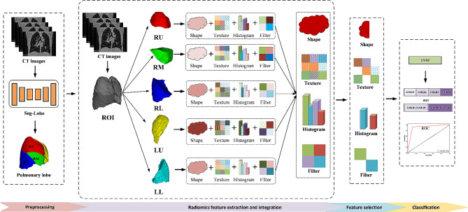
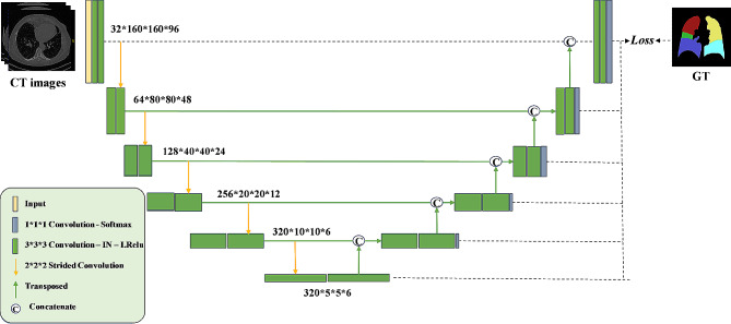
The retrospective study included 530 hospital patients. A lobe-based radiomics method was proposed to classify COPD severity using computed tomography (CT) images. First, we segmented the lung lobes with a convolutional neural network model. Secondly, the radiomic features of each lung lobe are extracted from CT images, the features of the five lung lobes are merged, and the selection of features is accomplished through the utilization of a variance threshold, t-Test, least absolute shrinkage and selection operator (LASSO). Finally, the COPD severity was classified by a support vector machine (SVM) classifier.
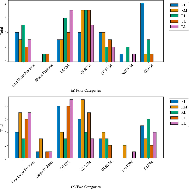
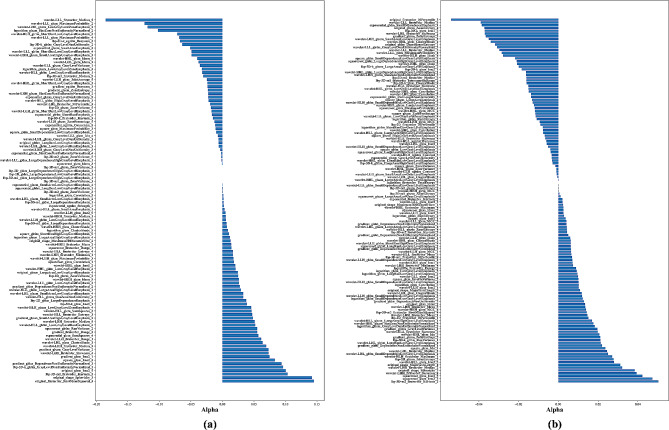
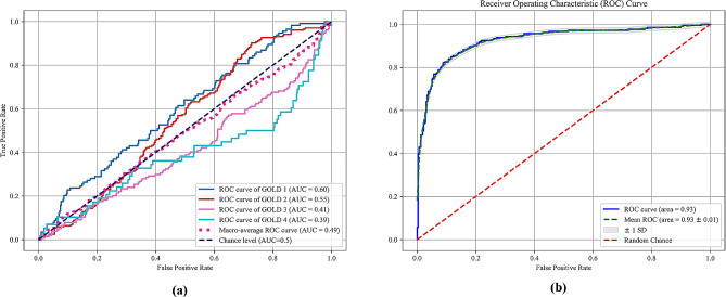
104 features were selected for staging COPD according to the Global initiative for chronic Obstructive Lung Disease (GOLD). The SVM classifier showed remarkable performance with an accuracy of 0.63. Moreover, an additional set of 132 features were selected to distinguish between milder (GOLD I + GOLD II) and more severe instances (GOLD III + GOLD IV) of COPD. The accuracy for SVM stood at 0.87.
The proposed method proved that the novel lobe-based radiomics method can significantly contribute to the refinement of COPD severity staging. By combining radiomic features from each lung lobe, it can obtain a more comprehensive and rich set of features and better capture the CT radiomic features of the lung than simply observing the lung as a whole.
慢性阻塞性肺疾病(COPD)是一种普遍且使人虚弱的呼吸系统疾病,在全球范围内造成了重大的医疗保健负担。准确分期 COPD 的严重程度对于患者管理和治疗计划至关重要。
本回顾性研究纳入了 530 名住院患者。提出了一种基于肺叶的放射组学方法,使用计算机断层扫描(CT)图像对 COPD 严重程度进行分类。首先,我们使用卷积神经网络模型对肺叶进行分割。其次,从 CT 图像中提取每个肺叶的放射组学特征,合并五个肺叶的特征,并通过使用方差阈值、t 检验、最小绝对收缩和选择算子(LASSO)选择特征。最后,使用支持向量机(SVM)分类器对 COPD 严重程度进行分类。
根据全球慢性阻塞性肺疾病倡议(GOLD),选择了 104 个特征对 COPD 进行分期。SVM 分类器的性能出色,准确率为 0.63。此外,还选择了另一组 132 个特征来区分更轻度(GOLD I + GOLD II)和更严重的 COPD 病例(GOLD III + GOLD IV)。SVM 的准确率为 0.87。
该研究提出的方法证明了基于肺叶的放射组学方法可以显著提高 COPD 严重程度分期的准确性。通过结合每个肺叶的放射组学特征,可以获得更全面、更丰富的特征集,并比单纯观察整个肺部更好地捕捉肺部的 CT 放射组学特征。