• 文献检索
  • 文档翻译
  • 深度研究
  • 学术资讯
  • Suppr Zotero 插件Zotero 插件
  • 邀请有礼
  • 套餐&价格
  • 历史记录
应用&插件
Suppr Zotero 插件Zotero 插件浏览器插件Mac 客户端Windows 客户端微信小程序
定价
高级版会员购买积分包购买API积分包
服务
文献检索文档翻译深度研究API 文档MCP 服务
关于我们
关于 Suppr公司介绍联系我们用户协议隐私条款
关注我们

Suppr 超能文献

核心技术专利:CN118964589B侵权必究
粤ICP备2023148730 号-1Suppr @ 2026

文献检索

告别复杂PubMed语法,用中文像聊天一样搜索,搜遍4000万医学文献。AI智能推荐,让科研检索更轻松。

立即免费搜索

文件翻译

保留排版,准确专业,支持PDF/Word/PPT等文件格式,支持 12+语言互译。

免费翻译文档

深度研究

AI帮你快速写综述,25分钟生成高质量综述,智能提取关键信息,辅助科研写作。

立即免费体验

有无对比纳米颗粒情况下胃癌细胞对宽带太赫兹辐射反应的统计分析

Statistical Analysis of Gastric Cancer Cells Response to Broadband Terahertz Radiation with and without Contrast Nanoparticles.

作者信息

Schreiner Oliver Daniel, Socotar Diana, Ciobanu Romeo Cristian, Schreiner Thomas Gabriel, Tamba Bogdan Ionel

机构信息

Department of Electrical Measurements and Materials, Gheorghe Asachi Technical University, 700050 Iasi, Romania.

CEMEX-Center for Experimental Medicine, "Grigore T. Popa" University of Medicine and Pharmacy, 700259 Iasi, Romania.

出版信息

Cancers (Basel). 2024 Jul 4;16(13):2454. doi: 10.3390/cancers16132454.

DOI:10.3390/cancers16132454
PMID:39001516
原文链接:https://pmc.ncbi.nlm.nih.gov/articles/PMC11240478/
Abstract

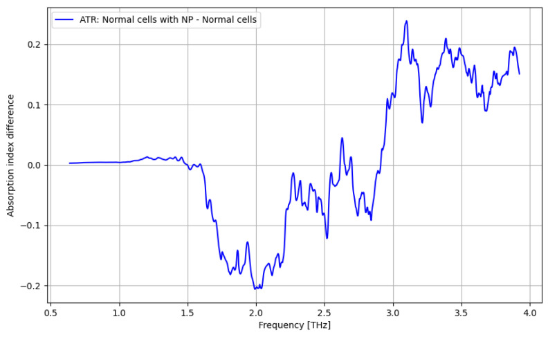
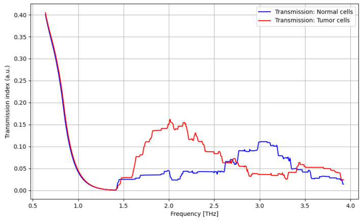
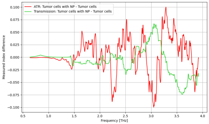
The paper describes the statistical analysis of the response of gastric cancer cells and normal cells to broadband terahertz radiation up to 4 THz, both with and without the use of nanostructured contrast agents. The THz spectroscopy analysis was comparatively performed under the ATR procedure and transmission measurement procedure. The statistical analysis was conducted towards multiple pairwise comparisons, including a support medium (without cells) versus a support medium with nanoparticles, normal cells versus normal cells with nanoparticles, and, respectively, tumor cells versus tumor cells with nanoparticles. When generally comparing the ATR procedure and transmission measurement procedure for a broader frequency domain, the differentiation between normal and tumor cells in the presence of contrast agents is superior when using the ATR procedure. THz contrast enhancement by using contrast agents derived from MRI-related contrast agents leads to only limited benefits and only for narrow THz frequency ranges, a disadvantage for THz medical imaging.

摘要

本文描述了胃癌细胞和正常细胞对高达4太赫兹的宽带太赫兹辐射的响应的统计分析,包括使用和不使用纳米结构造影剂的情况。太赫兹光谱分析是在衰减全反射(ATR)程序和透射测量程序下进行比较的。对多个成对比较进行了统计分析,包括支撑介质(无细胞)与含纳米颗粒的支撑介质、正常细胞与含纳米颗粒的正常细胞,以及肿瘤细胞与含纳米颗粒的肿瘤细胞。在更宽的频域中总体比较ATR程序和透射测量程序时,使用ATR程序时,在存在造影剂的情况下,正常细胞和肿瘤细胞之间的区分更优。使用源自磁共振成像(MRI)相关造影剂的造影剂实现的太赫兹对比度增强仅带来有限的益处,且仅适用于狭窄的太赫兹频率范围,这对太赫兹医学成像来说是一个缺点。

https://cdn.ncbi.nlm.nih.gov/pmc/blobs/c9b7/11240478/1eee4640e5e2/cancers-16-02454-g019.jpg
https://cdn.ncbi.nlm.nih.gov/pmc/blobs/c9b7/11240478/45402fa021dc/cancers-16-02454-g001.jpg
https://cdn.ncbi.nlm.nih.gov/pmc/blobs/c9b7/11240478/72dcfaed8f11/cancers-16-02454-g002.jpg
https://cdn.ncbi.nlm.nih.gov/pmc/blobs/c9b7/11240478/8a16fbb6ddbc/cancers-16-02454-g003.jpg
https://cdn.ncbi.nlm.nih.gov/pmc/blobs/c9b7/11240478/fac34a416a37/cancers-16-02454-g004.jpg
https://cdn.ncbi.nlm.nih.gov/pmc/blobs/c9b7/11240478/e5f84f7c65b2/cancers-16-02454-g005.jpg
https://cdn.ncbi.nlm.nih.gov/pmc/blobs/c9b7/11240478/a40651352ea7/cancers-16-02454-g006.jpg
https://cdn.ncbi.nlm.nih.gov/pmc/blobs/c9b7/11240478/c00615703d80/cancers-16-02454-g007.jpg
https://cdn.ncbi.nlm.nih.gov/pmc/blobs/c9b7/11240478/72fafd77b474/cancers-16-02454-g008.jpg
https://cdn.ncbi.nlm.nih.gov/pmc/blobs/c9b7/11240478/ab97f5d05102/cancers-16-02454-g009.jpg
https://cdn.ncbi.nlm.nih.gov/pmc/blobs/c9b7/11240478/6ce42b95c443/cancers-16-02454-g010.jpg
https://cdn.ncbi.nlm.nih.gov/pmc/blobs/c9b7/11240478/a523d0a7e3b6/cancers-16-02454-g011.jpg
https://cdn.ncbi.nlm.nih.gov/pmc/blobs/c9b7/11240478/43b79f3090ca/cancers-16-02454-g012.jpg
https://cdn.ncbi.nlm.nih.gov/pmc/blobs/c9b7/11240478/eca929234535/cancers-16-02454-g013.jpg
https://cdn.ncbi.nlm.nih.gov/pmc/blobs/c9b7/11240478/6f6c3eb06e72/cancers-16-02454-g014.jpg
https://cdn.ncbi.nlm.nih.gov/pmc/blobs/c9b7/11240478/c29ae1303c65/cancers-16-02454-g015.jpg
https://cdn.ncbi.nlm.nih.gov/pmc/blobs/c9b7/11240478/5a8d4199307d/cancers-16-02454-g016.jpg
https://cdn.ncbi.nlm.nih.gov/pmc/blobs/c9b7/11240478/ba8bcfa0c71d/cancers-16-02454-g017.jpg
https://cdn.ncbi.nlm.nih.gov/pmc/blobs/c9b7/11240478/2d513aafb676/cancers-16-02454-g018.jpg
https://cdn.ncbi.nlm.nih.gov/pmc/blobs/c9b7/11240478/1eee4640e5e2/cancers-16-02454-g019.jpg

相似文献

1
Statistical Analysis of Gastric Cancer Cells Response to Broadband Terahertz Radiation with and without Contrast Nanoparticles.有无对比纳米颗粒情况下胃癌细胞对宽带太赫兹辐射反应的统计分析
Cancers (Basel). 2024 Jul 4;16(13):2454. doi: 10.3390/cancers16132454.
2
Single-Shot Direct Transmission Terahertz Imaging Based on Intense Broadband Terahertz Radiation.基于强宽带太赫兹辐射的单次直接传输太赫兹成像
Sensors (Basel). 2024 Jun 26;24(13):4160. doi: 10.3390/s24134160.
3
[Progress of Detection Technology of Ultra-Broadband THz Time-Domain Spectroscopy].[超宽带太赫兹时域光谱检测技术进展]
Guang Pu Xue Yu Guang Pu Fen Xi. 2016 May;36(5):1277-83.
4
Broadband terahertz generation and detection at 10 nm scale.在 10nm 尺度上实现宽带太赫兹产生和探测。
Nano Lett. 2013 Jun 12;13(6):2884-8. doi: 10.1021/nl401219v. Epub 2013 May 23.
5
Clinical and experimental study of a terahertz time-domain system for the determination of the pathological margins of laryngeal carcinoma.用于确定喉癌病理边界的太赫兹时域系统的临床和实验研究。
World J Surg Oncol. 2022 Oct 12;20(1):339. doi: 10.1186/s12957-022-02788-8.
6
[The Detection of Ultra-Broadband Terahertz Spectroscopy of InP Wafer by Using Coherent Heterodyne Time-Domain Spectrometer].[利用相干外差时域光谱仪对磷化铟晶片进行超宽带太赫兹光谱检测]
Guang Pu Xue Yu Guang Pu Fen Xi. 2016 Feb;36(2):322-5.
7
Nanoscale biomaterials for terahertz imaging: A non-invasive approach for early cancer detection.用于太赫兹成像的纳米级生物材料:一种早期癌症检测的非侵入性方法。
Transl Oncol. 2023 Jan;27:101565. doi: 10.1016/j.tranon.2022.101565. Epub 2022 Nov 4.
8
Cancer detection in excised breast tumors using terahertz imaging and spectroscopy.利用太赫兹成像和光谱技术检测切除乳腺肿瘤中的癌症
Biomed Spectrosc Imaging. 2019;8(1-2):1-9. doi: 10.3233/bsi-190187. Epub 2019 Jul 9.
9
Terahertz imaging of excised oral cancer at frozen temperature.冷冻温度下切除的口腔癌的太赫兹成像
Biomed Opt Express. 2013 Jul 23;4(8):1413-21. doi: 10.1364/BOE.4.001413. eCollection 2013.
10
Terahertz broadband modulation in a biased BiFeO/Si heterojunction.偏置BiFeO/Si异质结中的太赫兹宽带调制
Opt Express. 2016 Nov 14;24(23):26618-26628. doi: 10.1364/OE.24.026618.

本文引用的文献

1
Terahertz Imaging and Spectroscopy in Cancer Diagnostics: A Technical Review.太赫兹成像与光谱技术在癌症诊断中的应用:技术综述
BME Front. 2020 Sep 25;2020:2547609. doi: 10.34133/2020/2547609. eCollection 2020.
2
High-throughput terahertz imaging: progress and challenges.高通量太赫兹成像:进展与挑战。
Light Sci Appl. 2023 Sep 15;12(1):233. doi: 10.1038/s41377-023-01278-0.
3
Optimal frequency determination for terahertz technology-based detection of colitis-related cancer in mice.基于太赫兹技术的结肠炎相关癌症在小鼠中检测的最佳频率确定。
J Biophotonics. 2023 Dec;16(12):e202300193. doi: 10.1002/jbio.202300193. Epub 2023 Aug 24.
4
Terahertz technology and its applications in head and neck diseases.太赫兹技术及其在头颈部疾病中的应用。
iScience. 2023 Jun 7;26(7):107060. doi: 10.1016/j.isci.2023.107060. eCollection 2023 Jul 21.
5
Radiographic and endoscopic screening to reduce gastric cancer mortality: a systematic review and meta-analysis.影像学和内镜筛查以降低胃癌死亡率:一项系统评价和荟萃分析。
Lancet Reg Health West Pac. 2023 Mar 11;35:100741. doi: 10.1016/j.lanwpc.2023.100741. eCollection 2023 Jun.
6
Super-resolution THz endoscope based on a hollow-core sapphire waveguide and a solid immersion lens.基于空芯蓝宝石波导和固体浸没透镜的太赫兹超分辨率内窥镜。
Opt Express. 2023 Apr 10;31(8):13366-13373. doi: 10.1364/OE.484650.
7
Revolutionary approaches for cancer diagnosis by terahertz-based spectroscopy and imaging.基于太赫兹光谱学和成像技术的癌症诊断创新方法。
Talanta. 2023 Jul 1;259:124483. doi: 10.1016/j.talanta.2023.124483. Epub 2023 Mar 29.
8
Epigenetic modification of gene expression in cancer cells by terahertz demethylation.太赫兹去甲基化对癌细胞中基因表达的表观遗传修饰。
Sci Rep. 2023 Mar 26;13(1):4930. doi: 10.1038/s41598-023-31828-w.
9
Nanoparticle-mediated cancer cell therapy: basic science to clinical applications.纳米颗粒介导的癌细胞治疗:从基础科学到临床应用。
Cancer Metastasis Rev. 2023 Sep;42(3):601-627. doi: 10.1007/s10555-023-10086-2. Epub 2023 Feb 24.
10
Early detection of gastric cancer high-resolution terahertz imaging system.早期胃癌检测 高分辨率太赫兹成像系统。
Front Bioeng Biotechnol. 2022 Dec 14;10:1052069. doi: 10.3389/fbioe.2022.1052069. eCollection 2022.