Verschuur Anouk S, Tax Chantal M W, Boomsma Martijn F, Carlson Helen L, van Wezel-Meijler Gerda, King Regan, Leemans Alexander, Leijser Lara M
Department of Radiology, Isala Hospital, Zwolle, Netherlands.
Image Sciences Institute, University Medical Center Utrecht, Utrecht, Netherlands.
Front Radiol. 2024 Jun 28;4:1416672. doi: 10.3389/fradi.2024.1416672. eCollection 2024.
The study aimed to (1) assess the feasibility constrained spherical deconvolution (CSD) tractography to reconstruct crossing fiber bundles with unsedated neonatal diffusion MRI (dMRI), and (2) demonstrate the impact of spatial and angular resolution and processing settings on tractography and derived quantitative measures.
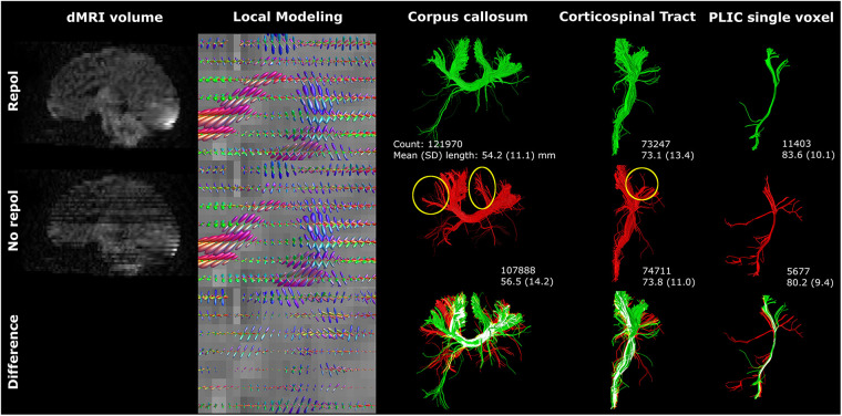
For the purpose of this study, the term-equivalent dMRIs (single-shell b800, and b2000, both 5 b0, and 45 gradient directions) of two moderate-late preterm infants (with and without motion artifacts) from a local cohort [Brain Imaging in Moderate-late Preterm infants (BIMP) study; Calgary, Canada] and one infant from the developing human connectome project with high-quality dMRI (using the b2600 shell, comprising 20 b0 and 128 gradient directions, from the multi-shell dataset) were selected. Diffusion tensor imaging (DTI) and CSD tractography were compared on b800 and b2000 dMRI. Varying image resolution modifications, (pre-)processing and tractography settings were tested to assess their impact on tractography. Each experiment involved visualizing local modeling and tractography for the corpus callosum and corticospinal tracts, and assessment of morphological and diffusion measures.
Contrary to DTI, CSD enabled reconstruction of crossing fibers. Tractography was susceptible to image resolution, (pre-) processing and tractography settings. In addition to visual variations, settings were found to affect streamline count, length, and diffusion measures (fractional anisotropy and mean diffusivity). Diffusion measures exhibited variations of up to 23%.
Reconstruction of crossing fiber bundles using CSD tractography with unsedated neonatal dMRI data is feasible. Tractography settings affected streamline reconstruction, warranting careful documentation of methods for reproducibility and comparison of cohorts.
本研究旨在(1)评估约束球形反卷积(CSD)纤维束成像在未镇静新生儿扩散磁共振成像(dMRI)中重建交叉纤维束的可行性,以及(2)证明空间和角度分辨率及处理设置对纤维束成像和衍生定量测量的影响。
为了本研究的目的,从本地队列[中晚期早产儿脑成像(BIMP)研究;加拿大卡尔加里]中选取了两名中晚期早产儿(有和没有运动伪影)的等效dMRI(单壳b800和b2000,均有5个b0和45个梯度方向),以及一名来自人类连接组计划的高质量dMRI的婴儿(使用来自多壳数据集的b2600壳,包括20个b0和128个梯度方向)。在b800和b2000 dMRI上比较扩散张量成像(DTI)和CSD纤维束成像。测试了不同的图像分辨率修改、(预)处理和纤维束成像设置,以评估它们对纤维束成像的影响。每个实验都包括可视化胼胝体和皮质脊髓束的局部建模和纤维束成像,以及形态学和扩散测量的评估。
与DTI相反,CSD能够重建交叉纤维。纤维束成像易受图像分辨率、(预)处理和纤维束成像设置的影响。除了视觉差异外,还发现这些设置会影响流线数量、长度和扩散测量(分数各向异性和平均扩散率)。扩散测量显示出高达23%的变化。
使用CSD纤维束成像和未镇静新生儿dMRI数据重建交叉纤维束是可行的。纤维束成像设置影响流线重建,因此需要仔细记录方法以确保可重复性和队列比较。