Fitrah Ahmad, Indriani R Vera, Hernowo Riza Putri Aulia, Nugraha Harry Galuh, Irawan M Naseh Sajadi Budi, Dewayani Birgitta Maria
Department of Radiology, Faculty of Medicine, University of Padjadjaran, Jl. Pasteur No.38, Pasteur, Sukajadi, Bandung City, West Java 40161, Indonesia.
Department of Orthopaedic and Traumatology, Faculty of Medicine, University of Padjadjaran, Jl. Pasteur No.38, Pasteur, Sukajadi, Bandung City, West Java 40161, Indonesia.
Radiol Case Rep. 2024 Jun 22;19(9):3833-3839. doi: 10.1016/j.radcr.2024.05.054. eCollection 2024 Sep.
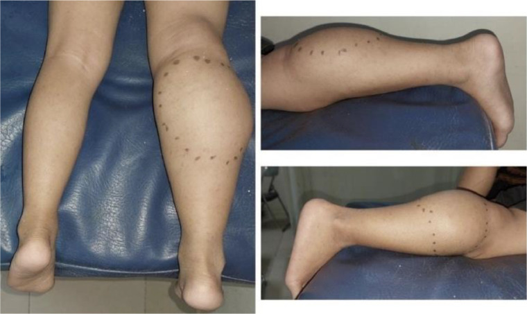
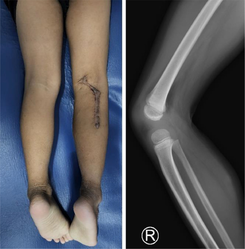
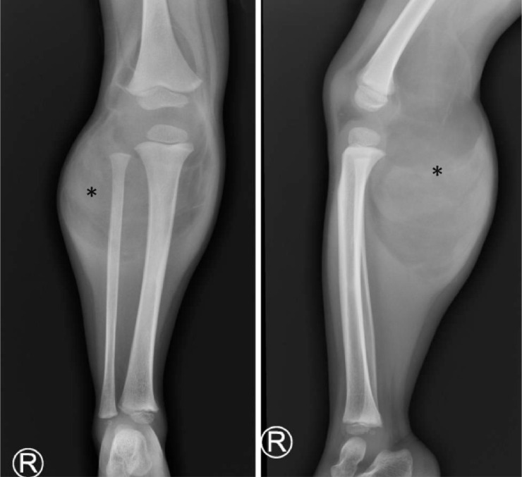
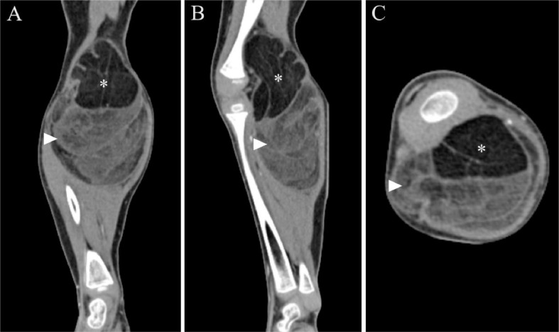
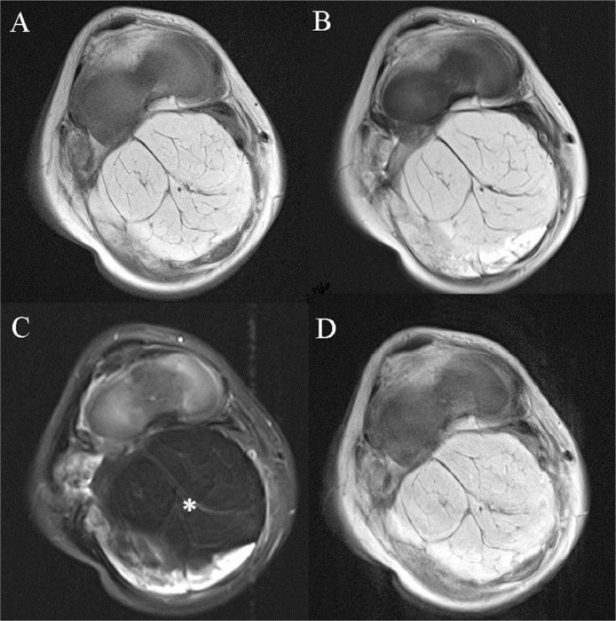
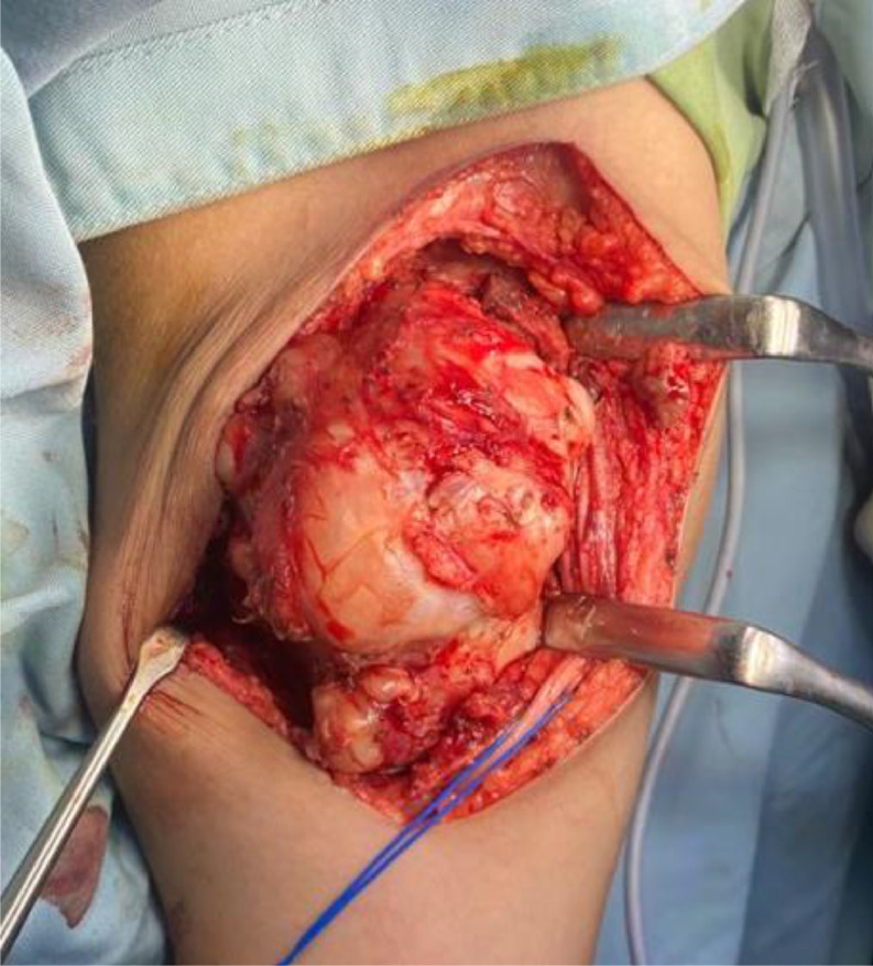
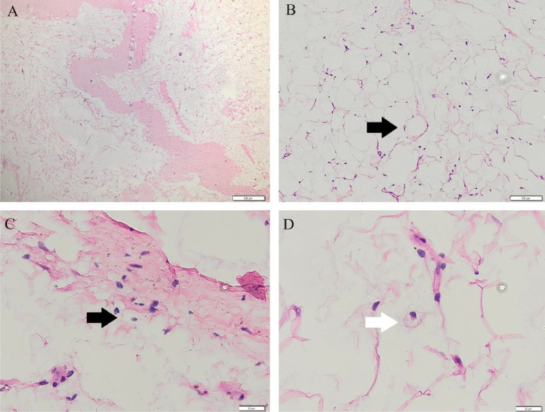
Lipoblastoma is a benign soft tissue tumor that originates from embryonic white fat. Lipoblastoma presents as a slow-growing mass that commonly occurs in the extremities of young children. Histological examination remains the gold standard in confirming lipoblastoma; however, radiology examination can help identify and evaluate the extent and characterization of the mass prior to the excision. Here, we report a 7-year-old male patient who presented with a painless mass in the right popliteal extending to the proximal cruris areas, and the imaging modalities suggested the presence of fat within the mass. The patient then underwent complete excision, and histopathology examination revealed lipoblastoma. This study highlights the possibility of lipoblastoma in older children and the role of imaging examinations in the diagnosis.
脂肪母细胞瘤是一种起源于胚胎白色脂肪的良性软组织肿瘤。脂肪母细胞瘤表现为生长缓慢的肿块,常见于幼儿的四肢。组织学检查仍是确诊脂肪母细胞瘤的金标准;然而,放射学检查有助于在切除术前识别和评估肿块的范围及特征。在此,我们报告一名7岁男性患者,其右腘窝出现无痛性肿块并延伸至近端小腿区域,影像学检查提示肿块内存在脂肪。该患者随后接受了完整切除,组织病理学检查显示为脂肪母细胞瘤。本研究强调了大龄儿童患脂肪母细胞瘤的可能性以及影像学检查在诊断中的作用。