• 文献检索
  • 文档翻译
  • 深度研究
  • 学术资讯
  • Suppr Zotero 插件Zotero 插件
  • 邀请有礼
  • 套餐&价格
  • 历史记录
应用&插件
Suppr Zotero 插件Zotero 插件浏览器插件Mac 客户端Windows 客户端微信小程序
定价
高级版会员购买积分包购买API积分包
服务
文献检索文档翻译深度研究API 文档MCP 服务
关于我们
关于 Suppr公司介绍联系我们用户协议隐私条款
关注我们

Suppr 超能文献

核心技术专利:CN118964589B侵权必究
粤ICP备2023148730 号-1Suppr @ 2026

文献检索

告别复杂PubMed语法,用中文像聊天一样搜索,搜遍4000万医学文献。AI智能推荐,让科研检索更轻松。

立即免费搜索

文件翻译

保留排版,准确专业,支持PDF/Word/PPT等文件格式,支持 12+语言互译。

免费翻译文档

深度研究

AI帮你快速写综述,25分钟生成高质量综述,智能提取关键信息,辅助科研写作。

立即免费体验

学龄儿童眼底视盘大小、颜色及杯盘比的纵向变化

Longitudinal changes of funduscopic optic disc size, color and cup-to-disc ratio in school children.

作者信息

Sameshima Seiji, Yamashita Takehiro, Terasaki Hiroto, Asaoka Ryo, Yoshihara Naoya, Kakiuchi Naoko, Sakamoto Taiji

机构信息

Department of Ophthalmology, Kagoshima University Graduate School of Medical and Dental Sciences, Kagoshima, Japan.

Department of Ophthalmology, Seirei Hamamatsu General Hospital, Shizuoka, Japan.

出版信息

Int J Retina Vitreous. 2024 Jul 25;10(1):51. doi: 10.1186/s40942-024-00570-4.

DOI:10.1186/s40942-024-00570-4
PMID:39054561
原文链接:https://pmc.ncbi.nlm.nih.gov/articles/PMC11270882/
Abstract

BACKGROUND

To investigate the relationship between changes in the optic disc size and color, cup-to-disc (C/D) ratio, and axial elongation in schoolchildren.

METHODS

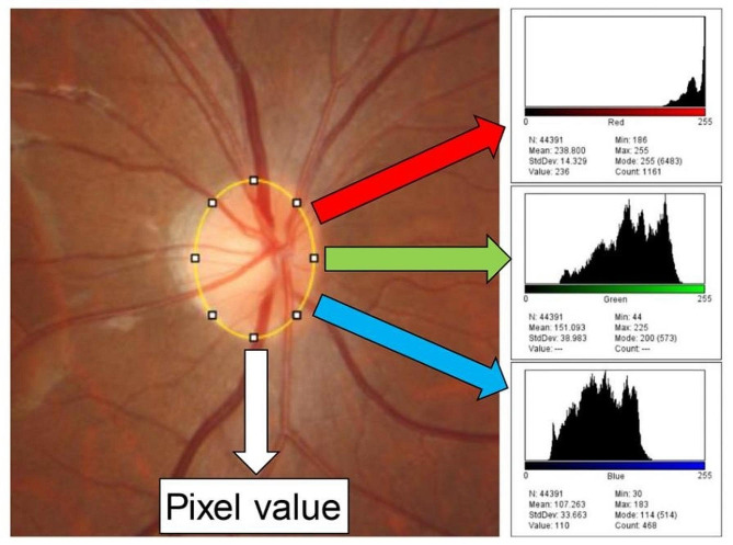
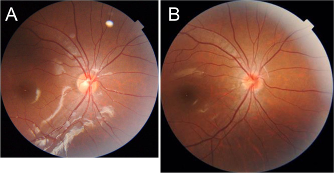
A prospective cohort study was performed in 75 right eyes of elementary school students for six years (from 8.5 to 14.5 years old). In the first and last year, all participants underwent optical axial length measurement and color fundus photography. The optic disc color was calculated by dividing the intensity of red by the sum of the intensity of red, green, and blue. The optic disc area was calculated by modifying the number of pixels according to Bennett's formula. The C/D ratio was calculated by dividing the vertical cup diameter by vertical optic disc diameter. Wilcoxon signed rank test was used to compare these optic disc parameters and axial length in the first and last year.

RESULTS

Mean axial length in the last year (24.82 mm) was significantly longer than that in the first year (23.34 mm). Likewise, the mean optic disc size was significantly smaller in the last year (41,946 pixels) than that in the first year (46,144 pixels). The mean optic disc color in the last year (0.49) was significantly more reddish than that in the first year (0.46), while the mean C/D ratio in last year (0.50) was significantly smaller than that in first year (0.52).

CONCLUSIONS

During the period from 8.5 years to 14.5 years of age, both the optic disc size and C/D ratio became smaller, while the color became more red.

摘要

背景

探讨学龄儿童视盘大小和颜色变化、杯盘比(C/D)与眼轴长度之间的关系。

方法

对75名小学生的右眼进行了为期六年(8.5至14.5岁)的前瞻性队列研究。在第一年和最后一年,所有参与者均接受了眼轴长度测量和彩色眼底照相。视盘颜色通过红色强度除以红色、绿色和蓝色强度之和来计算。视盘面积根据贝内特公式修改像素数量来计算。C/D比通过垂直杯径除以垂直视盘直径来计算。采用Wilcoxon符号秩检验比较第一年和最后一年的这些视盘参数和眼轴长度。

结果

最后一年的平均眼轴长度(24.82毫米)显著长于第一年(23.34毫米)。同样,最后一年的平均视盘大小(41,946像素)显著小于第一年(46,144像素)。最后一年的平均视盘颜色(0.49)比第一年(0.46)明显更红,而最后一年的平均C/D比(0.50)显著小于第一年(0.52)。

结论

在8.5岁至14.5岁期间,视盘大小和C/D比均变小,而颜色变得更红。

https://cdn.ncbi.nlm.nih.gov/pmc/blobs/6e2f/11270882/07a9ddcfe536/40942_2024_570_Fig2_HTML.jpg
https://cdn.ncbi.nlm.nih.gov/pmc/blobs/6e2f/11270882/185c0f3d8c36/40942_2024_570_Fig1_HTML.jpg
https://cdn.ncbi.nlm.nih.gov/pmc/blobs/6e2f/11270882/07a9ddcfe536/40942_2024_570_Fig2_HTML.jpg
https://cdn.ncbi.nlm.nih.gov/pmc/blobs/6e2f/11270882/185c0f3d8c36/40942_2024_570_Fig1_HTML.jpg
https://cdn.ncbi.nlm.nih.gov/pmc/blobs/6e2f/11270882/07a9ddcfe536/40942_2024_570_Fig2_HTML.jpg

相似文献

1
Longitudinal changes of funduscopic optic disc size, color and cup-to-disc ratio in school children.学龄儿童眼底视盘大小、颜色及杯盘比的纵向变化
Int J Retina Vitreous. 2024 Jul 25;10(1):51. doi: 10.1186/s40942-024-00570-4.
2
Birth weight and optic nerve head parameters.出生体重与视神经乳头参数。
Ophthalmology. 2009 Jun;116(6):1112-8. doi: 10.1016/j.ophtha.2008.12.061. Epub 2009 Apr 22.
3
[Morphological parameters of the posterior pole in a high myopia population aged over 40 years: a 10-year follow-up study].40岁以上高度近视人群后极部形态学参数:一项10年随访研究
Zhonghua Yan Ke Za Zhi. 2021 Dec 11;57(12):908-915. doi: 10.3760/cma.j.cn112142-20210125-00050.
4
Nonlinear behavior of certain optic nerve head parameters and their determinants in normal subjects.正常受试者中某些视神经乳头参数及其决定因素的非线性行为。
Ophthalmology. 2001 Dec;108(12):2358-68. doi: 10.1016/s0161-6420(01)00821-1.
5
Three-year changes in sex judgment using color fundus parameters in elementary school students.小学生眼底彩色参数性别判断的 3 年变化。
PLoS One. 2023 Nov 30;18(11):e0295123. doi: 10.1371/journal.pone.0295123. eCollection 2023.
6
Quantitative analysis of optic disc changes in school-age children with ametropia based on artificial intelligence.基于人工智能的学龄期屈光不正儿童视盘变化的定量分析
Int J Ophthalmol. 2023 Nov 18;16(11):1727-1733. doi: 10.18240/ijo.2023.11.01. eCollection 2023.
7
Quantification of peripapillary nerve fibre elevation and its association with axial length, optic disc tilt, and parapapillary atrophy area in young, healthy eyes.定量研究年轻健康眼的视盘旁神经纤维隆起及其与眼轴长度、视盘倾斜和视盘旁萎缩区的关系。
Eye (Lond). 2024 Apr;38(6):1112-1117. doi: 10.1038/s41433-023-02827-y. Epub 2023 Nov 15.
8
Measurement of optic nerve head parameters: comparison of optical coherence tomography with digital planimetry.视神经乳头参数的测量:光学相干断层扫描与数字平面测量法的比较
J Glaucoma. 2009 Oct-Nov;18(8):571-5. doi: 10.1097/IJG.0b013e3181996da6.
9
Optic disc measurements in myopia with optical coherence tomography and confocal scanning laser ophthalmoscopy.使用光学相干断层扫描和共焦扫描激光眼科显微镜对近视患者的视盘进行测量。
Invest Ophthalmol Vis Sci. 2007 Jul;48(7):3178-83. doi: 10.1167/iovs.06-1315.
10
Optic disc-fovea distance and myopia progression in school children: the Beijing Children Eye Study.视盘-黄斑中心凹距离与学龄儿童近视进展:北京儿童眼研究。
Acta Ophthalmol. 2018 Aug;96(5):e606-e613. doi: 10.1111/aos.13728. Epub 2018 Mar 25.

引用本文的文献

1
Longitudinal changes in the position and thickness of the peak peripapillary retinal nerve fiber layer in school children.学龄儿童视乳头周围视网膜神经纤维层峰值位置及厚度的纵向变化
Graefes Arch Clin Exp Ophthalmol. 2025 Mar 25. doi: 10.1007/s00417-025-06810-z.

本文引用的文献

1
Normative optical coherence tomography reference ranges of the optic nerve head, nerve fiber layer, and macula in healthy Saudi children.沙特健康儿童视神经头、神经纤维层和黄斑的正常光学相干断层扫描参考范围。
Saudi Med J. 2023 Nov 28;44(12):1269-1276. doi: 10.15537/smj.2023.44.12.20230517.
2
Optic Disc Measurements in Children by Optical Coherence Tomography.光学相干断层扫描技术在儿童视盘测量中的应用
J Glaucoma. 2023 May 1;32(5):361-368. doi: 10.1097/IJG.0000000000002159. Epub 2022 Nov 29.
3
Optic disc morphology in preterm children. Influence of gestational age and birth weight.
早产儿的视盘形态。胎龄和出生体重的影响。
J Fr Ophtalmol. 2021 Dec;44(10):1584-1588. doi: 10.1016/j.jfo.2021.03.009. Epub 2021 Oct 23.
4
Longitudinal Observation of Border Tissue Configuration During Axial Elongation in Childhood.儿童期轴向延长过程中边缘组织构型的纵向观察。
Invest Ophthalmol Vis Sci. 2021 Apr 1;62(4):10. doi: 10.1167/iovs.62.4.10.
5
Macular irregularities of optical coherence tomographic vertical cross sectional images in school age children.学龄儿童光学相干断层扫描垂直横截面图像的黄斑不规则性。
Sci Rep. 2021 Mar 5;11(1):5284. doi: 10.1038/s41598-021-84808-3.
6
The optic nerve head, lamina cribrosa, and nerve fiber layer in non-myopic and myopic children.非近视和近视儿童的视神经头、筛板和神经纤维层。
Exp Eye Res. 2020 Jun;195:108041. doi: 10.1016/j.exer.2020.108041. Epub 2020 Apr 28.
7
Disc Configuration as a Risk and Prognostic Factor in NAION: The Impact of Cup to Disc Ratio, Disc Diameter, and Crowding Index.视盘形态作为非动脉炎性前部缺血性视神经病变的风险和预后因素:杯盘比、视盘直径和拥挤指数的影响
Semin Ophthalmol. 2019;34(3):177-181. doi: 10.1080/08820538.2019.1620792. Epub 2019 Jun 4.
8
Global and regional estimates of prevalence of refractive errors: Systematic review and meta-analysis.屈光不正患病率的全球及区域估计:系统评价与荟萃分析
J Curr Ophthalmol. 2017 Sep 27;30(1):3-22. doi: 10.1016/j.joco.2017.08.009. eCollection 2018 Mar.
9
Optic Nerve Head Parameters Measured with Spectral-Domain Optical Coherence Tomography in Healthy Turkish Children: Normal Values, Repeatability, and Interocular Symmetry.在健康土耳其儿童中使用光谱域光学相干断层扫描测量的视神经乳头参数:正常值、重复性和双眼对称性。
Neuroophthalmology. 2017 Aug 8;42(2):83-89. doi: 10.1080/01658107.2017.1349806. eCollection 2018 Apr.
10
Changes in Axial Length and Progression of Visual Field Damage in Glaucoma.青光眼的眼轴长度变化与视野损害进展。
Invest Ophthalmol Vis Sci. 2018 Jan 1;59(1):407-417. doi: 10.1167/iovs.17-22949.