Cruz-Maya Iriczalli, Cirillo Valentina, Serrano-Bello Janeth, Serri Carla, Alvarez-Perez Marco Antonio, Guarino Vincenzo
Institute of Polymers, Composite and Biomaterials, National Research Council of Italy, Mostra d'Oltremare, V.le J.F.Kennedy 54, 80125 Naples, Italy.
Tissue Bioengineering Laboratory, Department of Posgraduate Studies and Research (DEPeI), School of Dentistry, Universidad Nacional Autonoma de Mexico (UNAM), Circuito Exterior s/n, Mexico City 04510, Mexico.
Pharmaceutics. 2024 Jul 11;16(7):925. doi: 10.3390/pharmaceutics16070925.
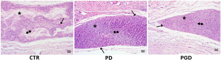
The use of electrospun fibers as anti-inflammatory drug carriers is currently one of the most interesting approaches for the design of drug delivery systems. In recent years, biodegradable polymers blended with naturally derived ones have been extensively studied to fabricate bioinspired platforms capable of driving biological responses by releasing selected molecular/pharmaceutical signals. Here, sodium diclofenac (DicNa)-loaded electrospun fibers, consisting of polycaprolactone (PCL) or gelatin-functionalized PCL, were studied to evaluate fibroblasts' in vitro and in vivo response. In vitro studies demonstrated that cell adhesion of L929 cells (≈70%) was not affected by the presence of DicNa after 4 h. Moreover, the initial burst release of the drug from PD and PGD fibers, e.g., 80 and 48%, respectively, after 5 h-combined with its sustained release-did not produce any cytotoxic effect and did not negatively influence the biological activity of the cells. In particular, it was demonstrated that the addition of gelatin concurred to slow down the release mechanism, thus limiting the antiproliferative effect of DicNa, as confirmed by the significant increase in cell viability and collagen deposition after 7 days, with respect to PCL alone. In vivo studies in a rat subcutaneous model also confirmed the ability of DicNa-loaded fibers to moderate the inflammatory/foreign body response independently through the presence of gelatin that played a significant role in supporting the formation of small-caliber vessels after 10 days of implantation. All of these results suggest using bicomponent fibers loaded with DicNa as a valid therapeutic tool capable of supporting the wound healing process and limiting in vivo inflammation and rejection phenomena.
将电纺纤维用作抗炎药物载体是目前药物递送系统设计中最具吸引力的方法之一。近年来,可生物降解聚合物与天然衍生聚合物的共混物已得到广泛研究,以构建能够通过释放选定的分子/药物信号来驱动生物学反应的仿生平台。在此,对负载双氯芬酸钠(DicNa)的电纺纤维进行了研究,该纤维由聚己内酯(PCL)或明胶功能化的PCL组成,以评估成纤维细胞的体外和体内反应。体外研究表明,4小时后,L929细胞的细胞黏附(约70%)不受DicNa存在的影响。此外,药物从PD和PGD纤维中的初始突释,例如5小时后分别为80%和48%,与其持续释放相结合,未产生任何细胞毒性作用,也未对细胞的生物学活性产生负面影响。特别是,已证明添加明胶有助于减缓释放机制,从而限制DicNa的抗增殖作用,这一点在7天后细胞活力和胶原蛋白沉积相对于单独的PCL显著增加中得到证实。大鼠皮下模型的体内研究也证实,负载DicNa的纤维能够独立地减轻炎症/异物反应,这是由于明胶的存在,在植入10天后,明胶在支持小口径血管形成方面发挥了重要作用。所有这些结果表明,使用负载DicNa的双组分纤维作为一种有效的治疗工具,能够支持伤口愈合过程并限制体内炎症和排斥现象。