Oral Pathology Department, Faculty of Oral and Dental Medicine, Misr International University, Cairo, Egypt.
Oral and Maxillofacial Pathology Department, Faculty of Dentistry, Cairo University, Cairo, Egypt.
BMC Oral Health. 2024 Aug 6;24(1):900. doi: 10.1186/s12903-024-04632-9.
Salivary gland neoplasms (SGNs) pose a challenge to both pathologists and clinicians. Despite research, the etiology of these neoplasms remains unclear. This study aimed to identify any potential association between the presence of hepatitis C virus (HCV) at the protein or gene level and epithelial salivary gland neoplasms.
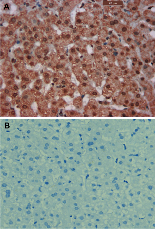
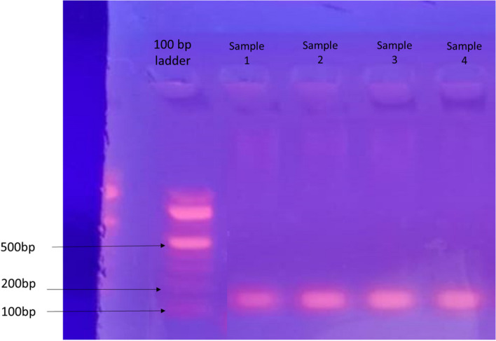
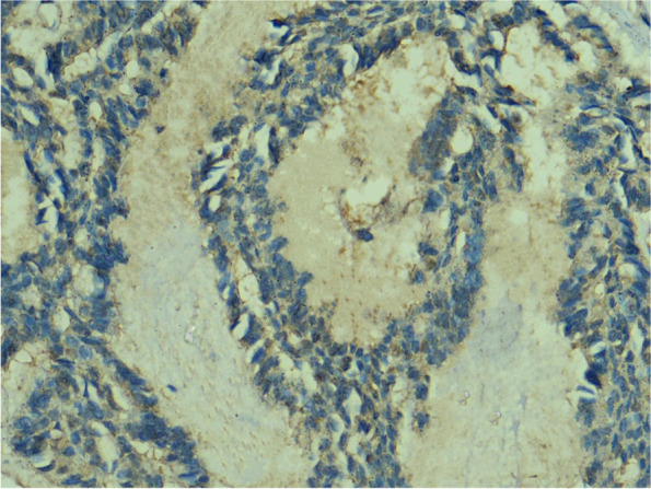
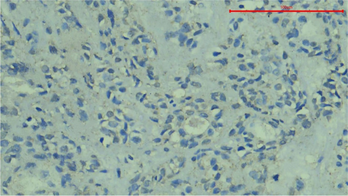
Formalin-fixed paraffin-embedded (FFPE) blocks of epithelial salivary gland neoplasms were retrieved from the archives of the Oral and Maxillofacial Pathology Department, Faculty of Dentistry, Cairo University within the 5-year period from 2016 to 2020. Immunohistochemistry was used to assess HCV core antigen, while reverse transcription polymerase chain reaction was employed for the evaluation of HCV RNA.
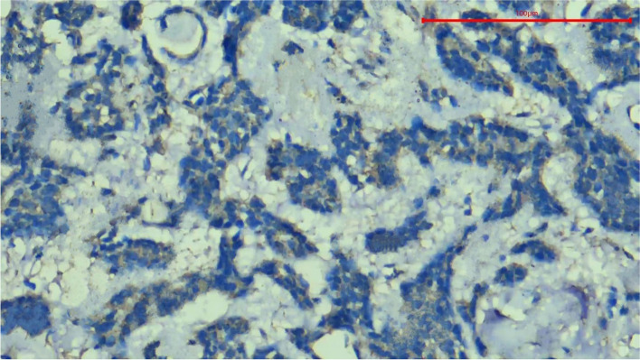
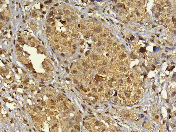
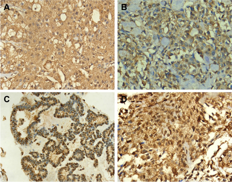
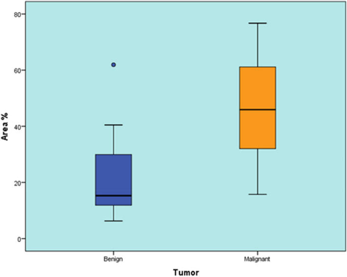
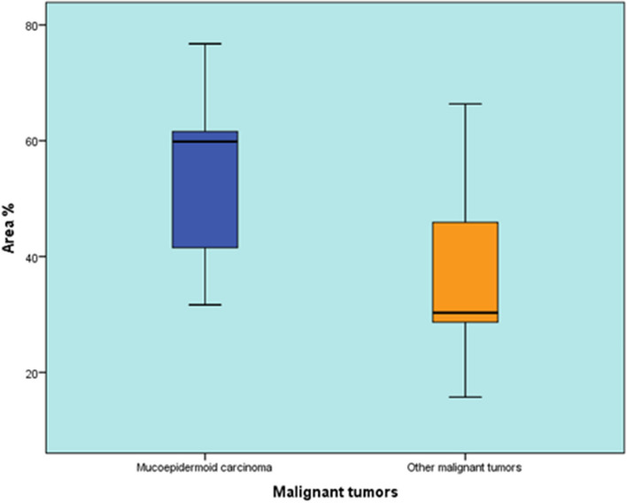
A total of 44 specimens were collected, 28 of which were benign neoplasms and 16 were malignant neoplasms. There was a statistically significant difference in HCV positivity between the two groups (P-value = 0.036). Benign tumors showed a statistically significant lower percentage of positive cases than malignant tumors. The localization of staining was also evaluated, revealing various patterns of HCV core antigen expression, including diffuse cytoplasmic, patchy cytoplasmic, nuclear, and a combination of nuclear and cytoplasmic expression. There was no statistically significant difference between the expression patterns in benign and malignant tumors (P-value = 0.616). Given that Pleomorphic Adenoma and Mucoepidermoid Carcinoma were the predominant tumor types in this study, four cases were selected for RNA detection. HCV RNA was detected in all cases using RT-PCR.
HCV core antigen is frequently detected in SGNs and is suggested to be a potential risk factor for the development of these neoplasms. Further studies are required to discover other biomarkers, their roles, and the pathways associated with HCV in SGNs.
唾液腺肿瘤(SGNs)对病理学家和临床医生都是一个挑战。尽管有研究,但这些肿瘤的病因仍不清楚。本研究旨在确定 HCV 蛋白或基因水平的存在与上皮性唾液腺肿瘤之间是否存在任何潜在关联。
从 2016 年至 2020 年的 5 年内,从开罗大学牙科学院口腔颌面病理学系的档案中检索上皮性唾液腺肿瘤的福尔马林固定石蜡包埋(FFPE)块。免疫组织化学用于评估 HCV 核心抗原,而逆转录聚合酶链反应用于评估 HCV RNA。
共收集了 44 个标本,其中 28 个为良性肿瘤,16 个为恶性肿瘤。两组之间 HCV 阳性率存在统计学差异(P 值=0.036)。良性肿瘤的阳性病例比例明显低于恶性肿瘤。还评估了染色的定位,显示 HCV 核心抗原表达存在各种模式,包括弥漫细胞质、斑片状细胞质、核和核质混合表达。良性和恶性肿瘤之间的表达模式无统计学差异(P 值=0.616)。鉴于多形性腺瘤和黏液表皮样癌是本研究中的主要肿瘤类型,因此选择了 4 例进行 RNA 检测。使用 RT-PCR 检测到所有病例均存在 HCV RNA。
HCV 核心抗原在上皮性唾液腺肿瘤中经常被检测到,提示其可能是这些肿瘤发生的潜在危险因素。需要进一步研究以发现其他生物标志物、它们的作用以及与 SGN 中 HCV 相关的途径。