• 文献检索
  • 文档翻译
  • 深度研究
  • 学术资讯
  • Suppr Zotero 插件Zotero 插件
  • 邀请有礼
  • 套餐&价格
  • 历史记录
应用&插件
Suppr Zotero 插件Zotero 插件浏览器插件Mac 客户端Windows 客户端微信小程序
定价
高级版会员购买积分包购买API积分包
服务
文献检索文档翻译深度研究API 文档MCP 服务
关于我们
关于 Suppr公司介绍联系我们用户协议隐私条款
关注我们

Suppr 超能文献

核心技术专利:CN118964589B侵权必究
粤ICP备2023148730 号-1Suppr @ 2026

文献检索

告别复杂PubMed语法,用中文像聊天一样搜索,搜遍4000万医学文献。AI智能推荐,让科研检索更轻松。

立即免费搜索

文件翻译

保留排版,准确专业,支持PDF/Word/PPT等文件格式,支持 12+语言互译。

免费翻译文档

深度研究

AI帮你快速写综述,25分钟生成高质量综述,智能提取关键信息,辅助科研写作。

立即免费体验

放大误差和眼轴长度对周边视网膜毛细血管密度和神经纤维层厚度的影响。

Effect of magnification error and axial length on circumpapillary capillary density and retinal nerve fiber layer thickness.

机构信息

Department of Ophthalmology, University of Tokyo Graduate School of Medicine, 7-3-1 Hongo, Bunkyo-ku, Tokyo, 113-8655, Japan.

Yotsuya Shirato Eye Clinic, 2-6 Samon-cho, Shinjuku-ku, Tokyo, Japan.

出版信息

Sci Rep. 2024 Aug 14;14(1):18874. doi: 10.1038/s41598-024-69864-9.

DOI:10.1038/s41598-024-69864-9
PMID:39143152
原文链接:https://pmc.ncbi.nlm.nih.gov/articles/PMC11324904/
Abstract

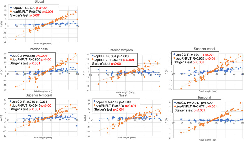
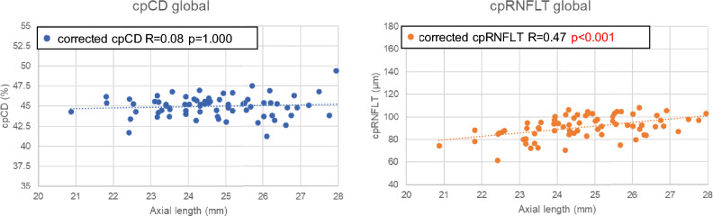
This study aimed to evaluate the effect of magnification error and axial length (AL) on circumpapillary capillary density (cpCD) and circumpapillary retinal nerve fiber layer thickness (cpRNFLT) in healthy eyes. Seventy-two healthy eyes of 72 subjects with AL 24.7 ± 1.5 mm (range: 20.9-28.0 mm) were enrolled in this retrospective cross-sectional study and underwent optical coherence tomography angiography scanning. Magnification corrected measurement areas were obtained using AL upon which corrected cpCD, cpRNFLT values were determined. Relationships between AL and the percentage difference between corrected and uncorrected values (ΔcpCD, ΔcpRNFLT) as well as the effect of AL on magnification corrected cpCD, cpRNFLT were evaluated. ΔcpCD significantly increased with AL in the global, inferior nasal and superior nasal sectors (all p < 0.001). ΔcpRNFLT significantly increased with AL in global and all sectors (all p < 0.001) and the correlations were significantly stronger than that of ΔcpCD-AL in all sectors (all p < 0.001). Corrected cpCD did not associate with AL while corrected cpRNFLT demonstrated a significant positive association with AL in the global (p = 0.005) and temporal sector (p < 0.001). Magnification error led to a significant underestimation of cpCD in eyes with longer AL although its underestimation and the effect of AL was smaller in comparison to that of cpRNFLT.

摘要

本研究旨在评估放大误差和眼轴(AL)对健康眼中的环周毛细血管密度(cpCD)和环周视网膜神经纤维层厚度(cpRNFLT)的影响。本回顾性横断面研究纳入了 72 名受试者的 72 只眼,AL 为 24.7±1.5mm(范围:20.9-28.0mm),并进行了光学相干断层扫描血管造影扫描。使用 AL 获取放大校正后的测量区域,确定校正后的 cpCD 和 cpRNFLT 值。评估了 AL 与校正和未校正值之间的百分比差异(ΔcpCD、ΔcpRNFLT)之间的关系以及 AL 对放大校正后的 cpCD 和 cpRNFLT 的影响。ΔcpCD 在全局、下鼻侧和上鼻侧区域均随 AL 增加而显著增加(均 p<0.001)。ΔcpRNFLT 在全局和所有区域均随 AL 增加而显著增加(均 p<0.001),且与所有区域的ΔcpCD-AL 相关性均显著更强(均 p<0.001)。校正后的 cpCD 与 AL 无关,而校正后的 cpRNFLT 与 AL 在全局(p=0.005)和颞侧区域(p<0.001)呈显著正相关。在 AL 较长的眼中,放大误差会导致 cpCD 显著低估,尽管与 cpRNFLT 相比,其低估和 AL 的影响较小。

https://cdn.ncbi.nlm.nih.gov/pmc/blobs/f495/11324904/5a644c932e3f/41598_2024_69864_Fig3_HTML.jpg
https://cdn.ncbi.nlm.nih.gov/pmc/blobs/f495/11324904/cbc95c216618/41598_2024_69864_Fig1_HTML.jpg
https://cdn.ncbi.nlm.nih.gov/pmc/blobs/f495/11324904/0d2475156224/41598_2024_69864_Fig2_HTML.jpg
https://cdn.ncbi.nlm.nih.gov/pmc/blobs/f495/11324904/5a644c932e3f/41598_2024_69864_Fig3_HTML.jpg
https://cdn.ncbi.nlm.nih.gov/pmc/blobs/f495/11324904/cbc95c216618/41598_2024_69864_Fig1_HTML.jpg
https://cdn.ncbi.nlm.nih.gov/pmc/blobs/f495/11324904/0d2475156224/41598_2024_69864_Fig2_HTML.jpg
https://cdn.ncbi.nlm.nih.gov/pmc/blobs/f495/11324904/5a644c932e3f/41598_2024_69864_Fig3_HTML.jpg

相似文献

1
Effect of magnification error and axial length on circumpapillary capillary density and retinal nerve fiber layer thickness.放大误差和眼轴长度对周边视网膜毛细血管密度和神经纤维层厚度的影响。
Sci Rep. 2024 Aug 14;14(1):18874. doi: 10.1038/s41598-024-69864-9.
2
Structure Function Relationships of Vessel Density and Retinal Nerve Fiber Layer Thickness in Early Glaucomatous Eyes With High Myopia.高度近视早期青光眼患者血管密度与视网膜神经纤维层厚度的结构-功能关系。
Invest Ophthalmol Vis Sci. 2023 Apr 3;64(4):14. doi: 10.1167/iovs.64.4.14.
3
Association Between Progressive Retinal Capillary Density Loss and Visual Field Progression in Open-Angle Glaucoma Patients According to Disease Stage.根据疾病阶段,广角型青光眼患者视网膜毛细血管密度进行性损失与视野进展之间的关系。
Am J Ophthalmol. 2021 Jun;226:137-147. doi: 10.1016/j.ajo.2021.01.015. Epub 2021 Jan 30.
4
Effects of axial length on retinal nerve fiber layer and macular ganglion cell-inner plexiform layer measured by spectral-domain OCT.轴向长度对通过频域光学相干断层扫描测量的视网膜神经纤维层和黄斑神经节细胞-内丛状层的影响。
Arq Bras Oftalmol. 2020 Aug;83(4):269-276. doi: 10.5935/0004-2749.20200039. Epub 2020 Jul 29.
5
Effect of refractive errors/axial length on peripapillary retinal nerve fibre layer thickness (RNFL) measured by Topcon SD-OCT.屈光不正/眼轴长度对Topcon SD-OCT测量的视盘周围视网膜神经纤维层厚度(RNFL)的影响。
J Pak Med Assoc. 2018 Jul;68(7):1054-1059.
6
Differences in Retinal Nerve Fiber Layer Thickness as Assessed on the Disc Center and Bruch's Membrane Opening Center in Myopic Eyes.近视眼中盘心和布鲁赫膜开口中心评估的视网膜神经纤维层厚度差异。
Ophthalmol Glaucoma. 2019 May-Jun;2(3):145-155. doi: 10.1016/j.ogla.2019.02.008. Epub 2019 Mar 2.
7
Measurements of OCT Angiography Complement OCT for Diagnosing Early Primary Open-Angle Glaucoma.OCT 血管造影测量对早期原发性开角型青光眼的诊断有 OCT 的补充作用。
Ophthalmol Glaucoma. 2022 May-Jun;5(3):262-274. doi: 10.1016/j.ogla.2021.09.012. Epub 2021 Oct 9.
8
Peripheral Ganglion Cell Complex Thickness and Retinal Microvasculature in Myopia Using Wide-Field Swept-Source OCT.应用超广角扫频源光学相干断层扫描测量近视的周边神经节细胞复合体厚度和视网膜微血管
Transl Vis Sci Technol. 2024 Aug 1;13(8):4. doi: 10.1167/tvst.13.8.4.
9
Circumpapillary microperimetry to detect glaucoma: a pilot study for sector-based comparison to circumpapillary retinal nerve fiber layer measurement.检测青光眼的视乳头周围微视野检查:一项基于象限与视乳头周围视网膜神经纤维层测量进行比较的初步研究。
Int Ophthalmol. 2019 Jan;39(1):127-136. doi: 10.1007/s10792-017-0796-8. Epub 2017 Dec 16.
10
Norms of Interocular Circumpapillary Retinal Nerve Fiber Layer Thickness Differences at 768 Retinal Locations.768个视网膜位置的双眼视盘周围视网膜神经纤维层厚度差异的规范
Transl Vis Sci Technol. 2020 Aug 12;9(9):23. doi: 10.1167/tvst.9.9.23. eCollection 2020 Aug.

引用本文的文献

1
Combined Probability Test for Sectoral Progression of the Circumpapillary Retinal Nerve Fiber Layer Thickness.视盘周围视网膜神经纤维层厚度扇形进展的联合概率检验
Transl Vis Sci Technol. 2025 Sep 2;14(9):4. doi: 10.1167/tvst.14.9.4.
2
Incidental detection of retinal nerve fiber layer thinning with slowed progression following optic nerve-carotid artery decompression: A case report.视神经-颈动脉减压术后视网膜神经纤维层变薄伴进展缓慢的偶然发现:一例报告。
Am J Ophthalmol Case Rep. 2025 Jul 8;39:102376. doi: 10.1016/j.ajoc.2025.102376. eCollection 2025 Sep.

本文引用的文献

1
Structure Function Relationships of Vessel Density and Retinal Nerve Fiber Layer Thickness in Early Glaucomatous Eyes With High Myopia.高度近视早期青光眼患者血管密度与视网膜神经纤维层厚度的结构-功能关系。
Invest Ophthalmol Vis Sci. 2023 Apr 3;64(4):14. doi: 10.1167/iovs.64.4.14.
2
Deep Optic Nerve Head Structures Associated With Increasing Axial Length in Healthy Myopic Eyes of Moderate Axial Length.健康中度近视眼轴增长相关的深层视神经头结构。
Am J Ophthalmol. 2023 May;249:156-166. doi: 10.1016/j.ajo.2023.01.003. Epub 2023 Jan 14.
3
Structure-Function Relationships and Glaucoma Detection with Magnification Correction of OCT Angiography.
光学相干断层扫描血管造影放大校正的结构-功能关系与青光眼检测
Ophthalmol Sci. 2022 Jan 30;2(2):100120. doi: 10.1016/j.xops.2022.100120. eCollection 2022 Jun.
4
Effects of optic nerve head-related parameters on retinal vessel calibers measurement results on fundus photographs.视盘相关参数对眼底照片视网膜血管直径测量结果的影响。
BMC Ophthalmol. 2022 May 12;22(1):215. doi: 10.1186/s12886-022-02428-5.
5
Diagnostic ability and sectoral structure-function relationship of circumpapillary and macular superficial vessel density in early glaucomatous eyes.早期青光眼眼中周边和黄斑浅层血管密度的诊断能力和节段性结构-功能关系。
Sci Rep. 2022 Apr 9;12(1):5991. doi: 10.1038/s41598-022-10033-1.
6
Macular and Optic Nerve Microvascular Alteration in Relation to Axial Length, by Optical Coherence Tomography Angiography (OCTA).光学相干断层扫描血管造影(OCTA)显示的黄斑和视神经微血管改变与眼轴长度的关系
Clin Ophthalmol. 2022 Mar 22;16:885-892. doi: 10.2147/OPTH.S354235. eCollection 2022.
7
Retinal microvasculature and optic disc alterations in non-pathological high myopia with optical coherence tomography angiography.光学相干断层扫描血管造影在病理性高度近视中的视网膜微血管和视盘改变。
Graefes Arch Clin Exp Ophthalmol. 2021 Nov;259(11):3221-3227. doi: 10.1007/s00417-021-05216-x. Epub 2021 May 7.
8
Induced Refractive Error Changes the Optical Coherence Tomography Angiography Transverse Magnification and Vascular Indices.诱导性屈光误差改变光学相干断层扫描血管造影的横向放大率和血管指数。
Am J Ophthalmol. 2021 Sep;229:230-241. doi: 10.1016/j.ajo.2021.04.012. Epub 2021 Apr 24.
9
OCT Angiography Artifacts in Glaucoma.青光眼的 OCT 血管造影伪影。
Ophthalmology. 2021 Oct;128(10):1426-1437. doi: 10.1016/j.ophtha.2021.03.036. Epub 2021 Apr 2.
10
Assessing the Use of Incorrectly Scaled Optical Coherence Tomography Angiography Images in Peer-Reviewed Studies: A Systematic Review.评估同行评审研究中使用错误缩放光学相干断层扫描血管造影图像的情况:系统评价。
JAMA Ophthalmol. 2020 Jan 1;138(1):86-94. doi: 10.1001/jamaophthalmol.2019.4821.