Yao Xiaohui, Zhu Yuan, Huang Zhenxing, Wang Yue, Cong Shan, Wan Liwen, Wu Ruodai, Chen Long, Hu Zhanli
Qingdao Innovation and Development Center, Harbin Engineering University, Qingdao, China.
Lauterbur Research Center for Biomedical Imaging, Shenzhen Institute of Advanced Technology, Chinese Academy of Sciences, Shenzhen, China.
Quant Imaging Med Surg. 2024 Aug 1;14(8):5460-5472. doi: 10.21037/qims-23-1028. Epub 2024 Jan 19.
Non-small cell lung cancer (NSCLC) patients with epidermal growth factor receptor-sensitizing (EGFR-sensitizing) mutations exhibit a positive response to tyrosine kinase inhibitors (TKIs). Given the limitations of current clinical predictive methods, it is critical to explore radiomics-based approaches. In this study, we leveraged deep-learning technology with multimodal radiomics data to more accurately predict EGFR-sensitizing mutations.
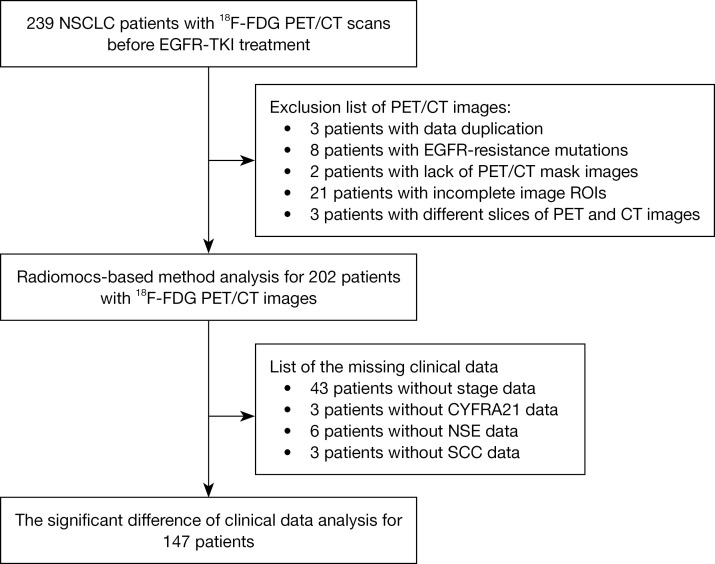
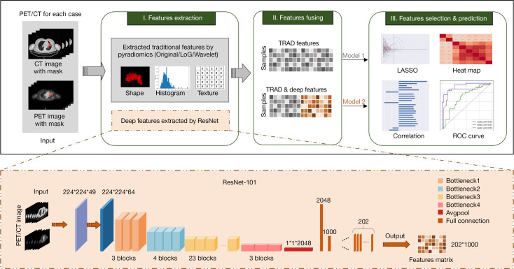
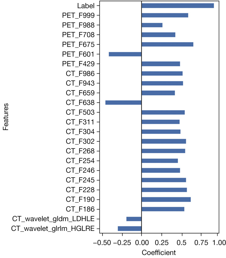
A total of 202 patients who underwent both flourine-18 fluorodeoxyglucose positron emission tomography/computed tomography (F-FDG PET/CT) scans and EGFR sequencing prior to treatment were included in this study. Deep and shallow features were extracted by a residual neural network and the Python package PyRadiomics, respectively. We used least absolute shrinkage and selection operator (LASSO) regression to select predictive features and applied a support vector machine (SVM) to classify the EGFR-sensitive patients. Moreover, we compared predictive performance across different deep models and imaging modalities.
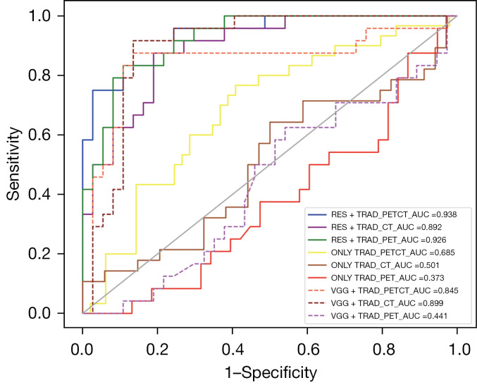
In the classification of EGFR-sensitive mutations, the areas under the curve (AUCs) of ResNet-based deep-shallow features and only shallow features from different multidata were as follows: RES_TRAD, PET/CT . CT-only . PET-only: 0.94 . 0.89 . 0.92; and ONLY_TRAD, PET/CT . CT-only . PET-only: 0.68 . 0.50 . 0.38. Additionally, the receiver operating characteristic (ROC) curves of the model using both deep and shallow features were significantly different from those of the model built using only shallow features (P<0.05).
Our findings suggest that deep features significantly enhance the detection of EGFR-sensitizing mutations, especially those extracted with ResNet. Moreover, PET/CT images are more effective than CT-only and PET-only images in producing EGFR-sensitizing mutation-related signatures.
表皮生长因子受体敏感(EGFR敏感)突变的非小细胞肺癌(NSCLC)患者对酪氨酸激酶抑制剂(TKIs)表现出阳性反应。鉴于当前临床预测方法的局限性,探索基于放射组学的方法至关重要。在本研究中,我们利用深度学习技术和多模态放射组学数据更准确地预测EGFR敏感突变。
本研究纳入了202例在治疗前接受过氟-18氟脱氧葡萄糖正电子发射断层扫描/计算机断层扫描(F-FDG PET/CT)和EGFR测序的患者。分别通过残差神经网络和Python包PyRadiomics提取深度和浅层特征。我们使用最小绝对收缩和选择算子(LASSO)回归选择预测特征,并应用支持向量机(SVM)对EGFR敏感患者进行分类。此外,我们比较了不同深度模型和成像模态的预测性能。
在EGFR敏感突变分类中,基于ResNet的深度-浅层特征以及来自不同多数据的仅浅层特征的曲线下面积(AUC)如下:RES_TRAD,PET/CT>仅CT>仅PET:0.94>0.89>0.92;以及ONLY_TRAD,PET/CT>仅CT>仅PET:0.68>0.50>0.38。此外,使用深度和浅层特征的模型的受试者操作特征(ROC)曲线与仅使用浅层特征构建的模型的ROC曲线有显著差异(P<0.05)。
我们的研究结果表明,深度特征显著增强了EGFR敏感突变的检测,尤其是用ResNet提取的特征。此外,PET/CT图像在生成与EGFR敏感突变相关的特征方面比仅CT图像和仅PET图像更有效。