Phour Jaison, Vassella Erik
Institute of Tissue Medicine & Pathology, University of Bern, 3008 Bern, Switzerland.
Biol Methods Protoc. 2024 Aug 22;9(1):bpae060. doi: 10.1093/biomethods/bpae060. eCollection 2024.
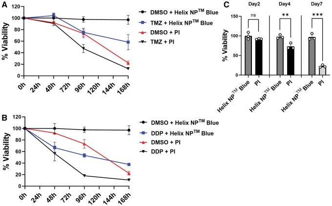
Spheroid cultures of cancer cell lines or primary cells represent a more clinically relevant model for predicting therapy response compared to two-dimensional cell culture. However, current live-dead staining protocols used for treatment response in spheroid cultures are often expensive, toxic to the cells, or limited in their ability to monitor therapy response over an extended period due to reduced stability. In our study, we have developed a cost-effective method utilizing calcein-AM and Helix NP™ Blue for live-dead staining, enabling the monitoring of therapy response of spheroid cultures for up to 10 days. Additionally, we used ICY BioImage Analysis and Z-stacks projection to calculate viability, which is a more accurate method for assessing treatment response compared to traditional methods on spheroid size. Using the example of glioblastoma cell lines and primary glioblastoma cells, we show that spheroid cultures typically exhibit a green outer layer of viable cells, a turquoise mantle of hypoxic quiescent cells, and a blue core of necrotic cells when visualized using confocal microscopy. Upon treatment of spheroids with the alkylating agent temozolomide, we observed a reduction in the viability of glioblastoma cells after an incubation period of 7 days. This method can also be adapted for monitoring therapy response in different cancer systems, offering a versatile and cost-effective approach for assessing therapy efficacy in three-dimensional culture models.
与二维细胞培养相比,癌细胞系或原代细胞的球体培养是一种更具临床相关性的预测治疗反应的模型。然而,目前用于球体培养中治疗反应的活死染色方案通常成本高昂、对细胞有毒,或者由于稳定性降低,在长时间监测治疗反应的能力方面存在局限性。在我们的研究中,我们开发了一种利用钙黄绿素-AM和Helix NP™ Blue进行活死染色的经济有效方法,能够监测球体培养长达10天的治疗反应。此外,我们使用ICY生物图像分析和Z-stack投影来计算存活率,与基于球体大小的传统方法相比,这是一种评估治疗反应更准确的方法。以胶质母细胞瘤细胞系和原发性胶质母细胞瘤细胞为例,我们发现,使用共聚焦显微镜观察时,球体培养通常显示出一层绿色的活细胞外层、一层蓝绿色的低氧静止细胞层和一个蓝色的坏死细胞核心。在用烷化剂替莫唑胺处理球体后,我们观察到胶质母细胞瘤细胞在孵育7天后存活率降低。该方法也可适用于监测不同癌症系统中的治疗反应,为评估三维培养模型中的治疗效果提供了一种通用且经济有效的方法。