• 文献检索
  • 文档翻译
  • 深度研究
  • 学术资讯
  • Suppr Zotero 插件Zotero 插件
  • 邀请有礼
  • 套餐&价格
  • 历史记录
应用&插件
Suppr Zotero 插件Zotero 插件浏览器插件Mac 客户端Windows 客户端微信小程序
定价
高级版会员购买积分包购买API积分包
服务
文献检索文档翻译深度研究API 文档MCP 服务
关于我们
关于 Suppr公司介绍联系我们用户协议隐私条款
关注我们

Suppr 超能文献

核心技术专利:CN118964589B侵权必究
粤ICP备2023148730 号-1Suppr @ 2026

文献检索

告别复杂PubMed语法,用中文像聊天一样搜索,搜遍4000万医学文献。AI智能推荐,让科研检索更轻松。

立即免费搜索

文件翻译

保留排版,准确专业,支持PDF/Word/PPT等文件格式,支持 12+语言互译。

免费翻译文档

深度研究

AI帮你快速写综述,25分钟生成高质量综述,智能提取关键信息,辅助科研写作。

立即免费体验

使用循环一致生成对抗网络结合注意力门控进行重建前处理以提高数字乳腺断层合成中的图像质量

Pre-Reconstruction Processing with the Cycle-Consist Generative Adversarial Network Combined with Attention Gate to Improve Image Quality in Digital Breast Tomosynthesis.

作者信息

Gomi Tsutomu, Ishihara Kotomi, Yamada Satoko, Koibuchi Yukio

机构信息

School of Allied Health Sciences, Kitasato University, Sagamihara 252-0373, Kanagawa, Japan.

Department of Radiology, NHO Takasaki General Medical Center, Takasaki 370-0829, Gunma, Japan.

出版信息

Diagnostics (Basel). 2024 Sep 4;14(17):1957. doi: 10.3390/diagnostics14171957.

DOI:10.3390/diagnostics14171957
PMID:39272741
原文链接:https://pmc.ncbi.nlm.nih.gov/articles/PMC11394014/
Abstract

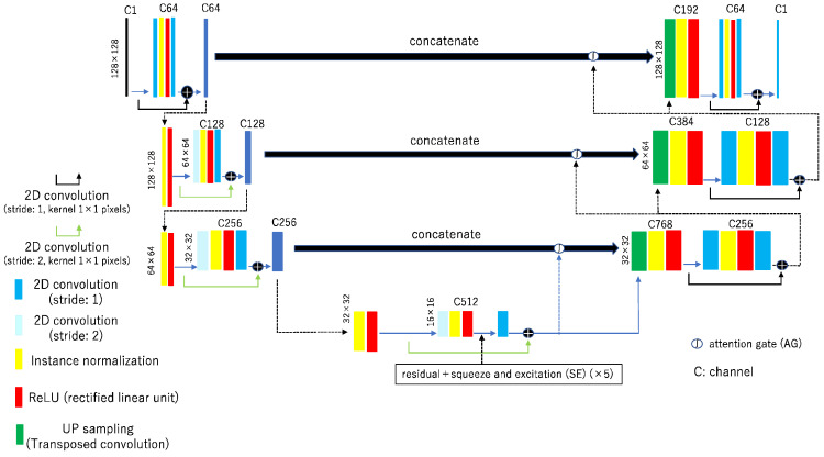
The current study proposed and evaluated "residual squeeze and excitation attention gate" (rSEAG), a novel network that can improve image quality by reducing distortion attributed to artifacts. This method was established by modifying the Cycle Generative Adversarial Network (cycleGAN)-based generator network using projection data for pre-reconstruction processing in digital breast tomosynthesis. Residual squeeze and excitation were installed in the bridge of the generator network, and the attention gate was installed in the skip connection between the encoder and decoder. Based on the radiation dose index (exposure index and division index) incident on the detector, the cases approved by the ethics committee and used for the study were classified as reference (675 projection images) and object (675 projection images). For the cases, unsupervised data containing a mixture of cases with and without masses were used. The cases were trained using cycleGAN with rSEAG and the conventional networks (ResUNet and U-Net). For testing, predictive processing was performed on cases (60 projection images) that were not used for learning. Images were generated using filtered backprojection reconstruction (kernel: Ramachandran and Lakshminarayanan) from projection data for testing data and without pre-reconstruction processing data (evaluation: in-focus plane). The distortion was evaluated using perception-based image quality evaluation (PIQE) analysis, texture analysis (feature: "Homogeneity" and "Contrast"), and a statistical model with a Gumbel distribution. PIQE has a low rSEAG value. Texture analysis showed that rSEAG and a network without cycleGAN were similar in terms of the "Contrast" feature. In dense breasts, ResUNet had the lowest "Contrast" feature and U-Net had differences between cases. The maximal variations in the Gumbel plot, rSEAG reduced the high-frequency ripple artifacts. In this study, rSEAG could improve distortion and reduce ripple artifacts.

摘要

当前的研究提出并评估了“残余挤压与激励注意力门”(rSEAG),这是一种新型网络,它可以通过减少由伪影引起的失真来提高图像质量。该方法是通过在数字乳腺断层合成中使用投影数据进行预重建处理,对基于循环生成对抗网络(cycleGAN)的生成器网络进行修改而建立的。在生成器网络的桥接部分安装了残余挤压与激励模块,在编码器和解码器之间的跳跃连接中安装了注意力门。根据入射到探测器上的辐射剂量指数(曝光指数和分割指数),伦理委员会批准并用于该研究的病例被分为参考组(675张投影图像)和对象组(675张投影图像)。对于这些病例,使用了包含有肿块和无肿块病例混合的无监督数据。使用带有rSEAG的cycleGAN和传统网络(ResUNet和U-Net)对这些病例进行训练。为了进行测试,对未用于学习的病例(60张投影图像)进行预测处理。使用滤波反投影重建(内核:拉马钱德兰和拉克什米纳拉亚南)从测试数据的投影数据生成图像,且不使用预重建处理数据(评估:聚焦平面)。使用基于感知的图像质量评估(PIQE)分析、纹理分析(特征:“均匀性”和“对比度”)以及具有耿贝尔分布的统计模型来评估失真。PIQE的rSEAG值较低。纹理分析表明,rSEAG和没有cycleGAN的网络在“对比度”特征方面相似。在致密乳腺中,ResUNet的“对比度”特征最低,U-Net在不同病例之间存在差异。在耿贝尔图中的最大变化方面,rSEAG减少了高频波纹伪影。在本研究中,rSEAG可以改善失真并减少波纹伪影。

https://cdn.ncbi.nlm.nih.gov/pmc/blobs/1bfa/11394014/3bbf0223a944/diagnostics-14-01957-g013.jpg
https://cdn.ncbi.nlm.nih.gov/pmc/blobs/1bfa/11394014/b5b6f591948b/diagnostics-14-01957-g001.jpg
https://cdn.ncbi.nlm.nih.gov/pmc/blobs/1bfa/11394014/758788c3a30a/diagnostics-14-01957-g002.jpg
https://cdn.ncbi.nlm.nih.gov/pmc/blobs/1bfa/11394014/c0341f6b0aa1/diagnostics-14-01957-g003.jpg
https://cdn.ncbi.nlm.nih.gov/pmc/blobs/1bfa/11394014/7a45f3d6453e/diagnostics-14-01957-g004.jpg
https://cdn.ncbi.nlm.nih.gov/pmc/blobs/1bfa/11394014/41427f3e046d/diagnostics-14-01957-g005a.jpg
https://cdn.ncbi.nlm.nih.gov/pmc/blobs/1bfa/11394014/4b0c2c730c3a/diagnostics-14-01957-g006.jpg
https://cdn.ncbi.nlm.nih.gov/pmc/blobs/1bfa/11394014/414828426361/diagnostics-14-01957-g007.jpg
https://cdn.ncbi.nlm.nih.gov/pmc/blobs/1bfa/11394014/39f5ca352c82/diagnostics-14-01957-g008.jpg
https://cdn.ncbi.nlm.nih.gov/pmc/blobs/1bfa/11394014/a3f4aaac1bd4/diagnostics-14-01957-g009.jpg
https://cdn.ncbi.nlm.nih.gov/pmc/blobs/1bfa/11394014/de1af63b5715/diagnostics-14-01957-g010.jpg
https://cdn.ncbi.nlm.nih.gov/pmc/blobs/1bfa/11394014/a85e080acf5d/diagnostics-14-01957-g011.jpg
https://cdn.ncbi.nlm.nih.gov/pmc/blobs/1bfa/11394014/ae1e0826dc6b/diagnostics-14-01957-g012.jpg
https://cdn.ncbi.nlm.nih.gov/pmc/blobs/1bfa/11394014/3bbf0223a944/diagnostics-14-01957-g013.jpg

相似文献

1
Pre-Reconstruction Processing with the Cycle-Consist Generative Adversarial Network Combined with Attention Gate to Improve Image Quality in Digital Breast Tomosynthesis.使用循环一致生成对抗网络结合注意力门控进行重建前处理以提高数字乳腺断层合成中的图像质量
Diagnostics (Basel). 2024 Sep 4;14(17):1957. doi: 10.3390/diagnostics14171957.
2
Evaluation of a Generative Adversarial Network to Improve Image Quality and Reduce Radiation-Dose during Digital Breast Tomosynthesis.评估生成对抗网络以提高数字乳腺断层合成期间的图像质量并降低辐射剂量
Diagnostics (Basel). 2022 Feb 14;12(2):495. doi: 10.3390/diagnostics12020495.
3
Improved digital chest tomosynthesis image quality by use of a projection-based dual-energy virtual monochromatic convolutional neural network with super resolution.基于投影的双能虚拟单能量卷积神经网络与超分辨技术改善数字胸部断层合成图像质量
PLoS One. 2020 Dec 31;15(12):e0244745. doi: 10.1371/journal.pone.0244745. eCollection 2020.
4
A two-step method to improve image quality of CBCT with phantom-based supervised and patient-based unsupervised learning strategies.基于体模的有监督和基于患者的无监督学习策略两步法提高 CBCT 图像质量。
Phys Med Biol. 2022 Apr 11;67(8). doi: 10.1088/1361-6560/ac6289.
5
Normalization of HE-stained histological images using cycle consistent generative adversarial networks.使用循环一致生成对抗网络对 HE 染色组织学图像进行归一化。
Diagn Pathol. 2021 Aug 6;16(1):71. doi: 10.1186/s13000-021-01126-y.
6
Usefulness of a Metal Artifact Reduction Algorithm in Digital Tomosynthesis Using a Combination of Hybrid Generative Adversarial Networks.使用混合生成对抗网络组合的数字断层合成中金属伪影减少算法的效用
Diagnostics (Basel). 2021 Sep 6;11(9):1629. doi: 10.3390/diagnostics11091629.
7
A novel pre-processing technique for improving image quality in digital breast tomosynthesis.一种用于提高数字乳腺断层合成图像质量的新型预处理技术。
Med Phys. 2017 Feb;44(2):417-425. doi: 10.1002/mp.12078. Epub 2017 Feb 2.
8
Multi-scale cascaded networks for synthesis of mammogram to decrease intensity distortion and increase model-based perceptual similarity.多尺度级联网络用于合成乳腺 X 线照片,以降低强度失真并提高基于模型的感知相似性。
Med Phys. 2023 Feb;50(2):837-853. doi: 10.1002/mp.16007. Epub 2022 Oct 24.
9
Low-light image enhancement using generative adversarial networks.使用生成对抗网络的低光照图像增强
Sci Rep. 2024 Aug 9;14(1):18489. doi: 10.1038/s41598-024-69505-1.
10
Nighttime road scene image enhancement based on cycle-consistent generative adversarial network.基于循环一致生成对抗网络的夜间道路场景图像增强
Sci Rep. 2024 Jun 22;14(1):14375. doi: 10.1038/s41598-024-65270-3.

本文引用的文献

1
Motion artifact correction in cardiac CT using cross-phase temporospatial information and synergistic attention gate and spatial transformer sub-networks.利用交叉相位时空间信息以及协同注意门和空间变换子网络校正心脏 CT 中的运动伪影。
Phys Med Biol. 2024 Feb 2;69(3). doi: 10.1088/1361-6560/ad1b6a.
2
Improvement of 2D cine image quality using 3D priors and cycle generative adversarial network for low field MRI-guided radiation therapy.利用三维先验信息和循环生成对抗网络改善低场 MRI 引导放射治疗的二维电影图像质量。
Med Phys. 2024 May;51(5):3495-3509. doi: 10.1002/mp.16860. Epub 2023 Dec 3.
3
Improving portable low-field MRI image quality through image-to-image translation using paired low- and high-field images.
通过使用配对的低场和高场图像进行图像到图像的转换来提高便携式低场 MRI 图像质量。
Sci Rep. 2023 Dec 1;13(1):21183. doi: 10.1038/s41598-023-48438-1.
4
A lightweight multi-modality medical image semantic segmentation network base on the novel UNeXt and Wave-MLP.基于新型 UNeXt 和 Wave-MLP 的轻量级多模态医学图像语义分割网络。
Comput Med Imaging Graph. 2024 Jan;111:102311. doi: 10.1016/j.compmedimag.2023.102311. Epub 2023 Nov 8.
5
Visual and quantitative evaluation of microcalcifications in mammograms with deep learning-based super-resolution.基于深度学习的超分辨率技术在乳腺 X 光片中微钙化的可视化和定量评估。
Eur J Radiol. 2022 Sep;154:110433. doi: 10.1016/j.ejrad.2022.110433. Epub 2022 Jul 6.
6
Evaluation of a Generative Adversarial Network to Improve Image Quality and Reduce Radiation-Dose during Digital Breast Tomosynthesis.评估生成对抗网络以提高数字乳腺断层合成期间的图像质量并降低辐射剂量
Diagnostics (Basel). 2022 Feb 14;12(2):495. doi: 10.3390/diagnostics12020495.
7
A Data Set and Deep Learning Algorithm for the Detection of Masses and Architectural Distortions in Digital Breast Tomosynthesis Images.用于数字乳腺断层合成图像中肿块和结构扭曲检测的数据集合和深度学习算法。
JAMA Netw Open. 2021 Aug 2;4(8):e2119100. doi: 10.1001/jamanetworkopen.2021.19100.
8
Impact of Artificial Intelligence Decision Support Using Deep Learning on Breast Cancer Screening Interpretation with Single-View Wide-Angle Digital Breast Tomosynthesis.深度学习辅助人工智能决策对单视图广角数字乳腺断层合成乳腺癌筛查解读的影响。
Radiology. 2021 Sep;300(3):529-536. doi: 10.1148/radiol.2021204432. Epub 2021 Jul 6.
9
Synthesis of Mammogram From Digital Breast Tomosynthesis Using Deep Convolutional Neural Network With Gradient Guided cGANs.基于深度卷积神经网络和梯度引导 cGANs 的乳腺断层合成数字乳腺 X 线摄影术
IEEE Trans Med Imaging. 2021 Aug;40(8):2080-2091. doi: 10.1109/TMI.2021.3071544. Epub 2021 Jul 30.
10
Deep Convolutional Neural Network With Adversarial Training for Denoising Digital Breast Tomosynthesis Images.基于对抗训练的深度卷积神经网络用于数字乳腺断层合成图像去噪。
IEEE Trans Med Imaging. 2021 Jul;40(7):1805-1816. doi: 10.1109/TMI.2021.3066896. Epub 2021 Jun 30.