• 文献检索
  • 文档翻译
  • 深度研究
  • 学术资讯
  • Suppr Zotero 插件Zotero 插件
  • 邀请有礼
  • 套餐&价格
  • 历史记录
应用&插件
Suppr Zotero 插件Zotero 插件浏览器插件Mac 客户端Windows 客户端微信小程序
定价
高级版会员购买积分包购买API积分包
服务
文献检索文档翻译深度研究API 文档MCP 服务
关于我们
关于 Suppr公司介绍联系我们用户协议隐私条款
关注我们

Suppr 超能文献

核心技术专利:CN118964589B侵权必究
粤ICP备2023148730 号-1Suppr @ 2026

文献检索

告别复杂PubMed语法,用中文像聊天一样搜索,搜遍4000万医学文献。AI智能推荐,让科研检索更轻松。

立即免费搜索

文件翻译

保留排版,准确专业,支持PDF/Word/PPT等文件格式,支持 12+语言互译。

免费翻译文档

深度研究

AI帮你快速写综述,25分钟生成高质量综述,智能提取关键信息,辅助科研写作。

立即免费体验

金属纳米粒子在结石形成中的作用。

The Role of Metal Nanoparticles in the Pathogenesis of Stone Formation.

机构信息

Department of Surgical Dentistry and Implantology of the N.D. Yushchuk Institute of Continuing Professional Education, Russian University of Medicine of the Ministry of Health of the Russian Federation, 4 Dolgorukovskaya St., 127006 Moscow, Russia.

National Research Centre "Kurchatov Institute", 59 Leninskiy Prospekt, 119333 Moscow, Russia.

出版信息

Int J Mol Sci. 2024 Sep 5;25(17):9609. doi: 10.3390/ijms25179609.

DOI:10.3390/ijms25179609
PMID:39273555
原文链接:https://pmc.ncbi.nlm.nih.gov/articles/PMC11394863/
Abstract

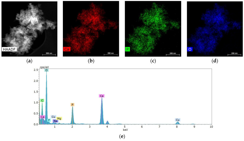
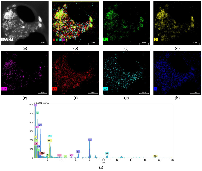
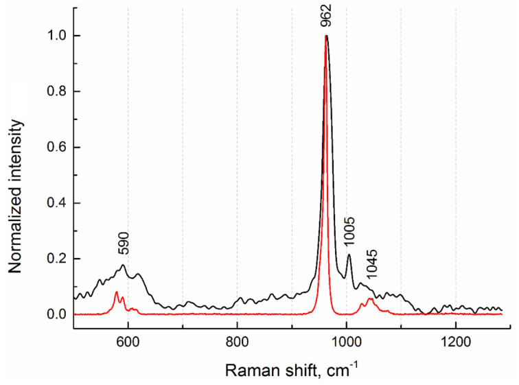
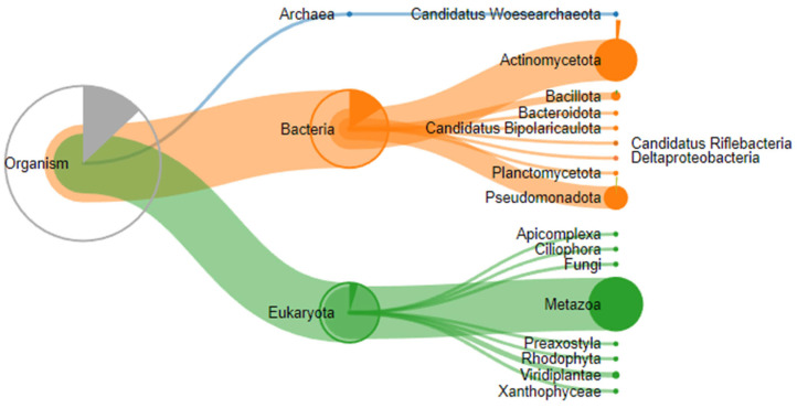
The process of stone formation in the human body remains incompletely understood, which requires clinical and laboratory studies and the formulation of a new endogenous, nanotechnological concept of the mechanism of origin and formation of crystallization centers. Previously, the mechanism of sialolithiasis was considered a congenital disease associated with the pathology of the ducts in the structure of the glands themselves. To date, such morphological changes of congenital nature can be considered from the position of the intrauterine formation of endogenous bacterial infections complicated by the migration of antigenic structures initiating the formation of crystallization centers. The present work is devoted to the study of the morphology and composition of stones obtained as a result of surgical interventions for sialolithiasis. Presumably, nanoparticles of metals and other chemical compounds can be structural components of crystallization centers or incorporated into the conditions of chronic endogenous inflammation and the composition of antigenic structures, in complexes with protein and bacterial components. X-ray microtomography, X-ray fluorescence analysis, scanning transmission electron microscopy and microanalysis, mass spectrometry, and Raman spectroscopy were used to study the pathogenesis of stone formation. Immunoglobulins (Igs) of classes A and G, as well as nanoparticles of metals Pb, Fe, Cr, and Mo, were found in the internal structure of the stones. The complex of antigenic structures was an ovoid calcified layered matrix of polyvid microbial biofilms, with the inclusion of metal nanoparticles and chemical elements, as well as immunoglobulins. The obtained results of clinical and laboratory studies allow us to broaden the view on the pathogenesis of stone formation and suggest that the occurrence of the calcification of antigenic structures may be associated with the formation of IgG4-associated disease.

摘要

人体结石形成的过程仍不完全清楚,这需要临床和实验室研究,并提出新的内源性、纳米技术概念来解释结晶中心的起源和形成机制。以前,涎石病的发病机制被认为是一种与腺体自身结构中的导管病理学相关的先天性疾病。迄今为止,这种先天性的形态学变化可以从内生细菌感染的宫内形成的角度来考虑,这种感染会引发抗原结构的迁移,从而启动结晶中心的形成。本研究致力于研究外科手术治疗涎石病所获得的结石的形态和成分。推测金属和其他化合物的纳米颗粒可能是结晶中心的结构成分,或被纳入慢性内源性炎症和抗原结构的组成中,与蛋白质和细菌成分形成复合物。本研究采用 X 射线微断层扫描、X 射线荧光分析、扫描透射电子显微镜和微分析、质谱和拉曼光谱法来研究结石形成的发病机制。在结石的内部结构中发现了 A 类和 G 类免疫球蛋白(Igs)以及 Pb、Fe、Cr 和 Mo 等金属的纳米颗粒。抗原结构复合物是多微生物生物膜的卵形钙化层状基质,包含金属纳米颗粒和化学元素以及免疫球蛋白。临床和实验室研究的结果使我们能够更全面地了解结石形成的发病机制,并提示抗原结构钙化的发生可能与 IgG4 相关疾病的形成有关。

https://cdn.ncbi.nlm.nih.gov/pmc/blobs/2af2/11394863/3e53c4697c21/ijms-25-09609-g010.jpg
https://cdn.ncbi.nlm.nih.gov/pmc/blobs/2af2/11394863/559427e34ce2/ijms-25-09609-g001.jpg
https://cdn.ncbi.nlm.nih.gov/pmc/blobs/2af2/11394863/0615ae3daf4d/ijms-25-09609-g002.jpg
https://cdn.ncbi.nlm.nih.gov/pmc/blobs/2af2/11394863/88a2a1fbc730/ijms-25-09609-g003.jpg
https://cdn.ncbi.nlm.nih.gov/pmc/blobs/2af2/11394863/c2843060fa74/ijms-25-09609-g004.jpg
https://cdn.ncbi.nlm.nih.gov/pmc/blobs/2af2/11394863/36e213c2fc57/ijms-25-09609-g005.jpg
https://cdn.ncbi.nlm.nih.gov/pmc/blobs/2af2/11394863/c624489dc04c/ijms-25-09609-g006.jpg
https://cdn.ncbi.nlm.nih.gov/pmc/blobs/2af2/11394863/431f74335b26/ijms-25-09609-g007.jpg
https://cdn.ncbi.nlm.nih.gov/pmc/blobs/2af2/11394863/c8d9598a523e/ijms-25-09609-g008.jpg
https://cdn.ncbi.nlm.nih.gov/pmc/blobs/2af2/11394863/2885ef2d8e22/ijms-25-09609-g009.jpg
https://cdn.ncbi.nlm.nih.gov/pmc/blobs/2af2/11394863/3e53c4697c21/ijms-25-09609-g010.jpg

相似文献

1
The Role of Metal Nanoparticles in the Pathogenesis of Stone Formation.金属纳米粒子在结石形成中的作用。
Int J Mol Sci. 2024 Sep 5;25(17):9609. doi: 10.3390/ijms25179609.
2
Possible etiology of calculi formation in salivary glands: biophysical analysis of calculus.涎腺结石形成的可能病因:结石的生物物理分析
Med Mol Morphol. 2005 Sep;38(3):189-95. doi: 10.1007/s00795-005-0290-7.
3
Bacterial Biofilm in Salivary Gland Stones: Cause or Consequence?唾液腺结石中的细菌生物膜:原因还是结果?
Otolaryngol Head Neck Surg. 2016 Mar;154(3):449-53. doi: 10.1177/0194599815622425. Epub 2016 Jan 12.
4
Neutrophil Extracellular Traps Promote the Development and Growth of Human Salivary Stones.中性粒细胞胞外诱捕网促进人唾液结石的发生和生长。
Cells. 2020 Sep 22;9(9):2139. doi: 10.3390/cells9092139.
5
Ultrastructural analysis of the submandibular sialoliths: Raman spectroscopy and electron back-scatter studies.下颌下涎石的超微结构分析:拉曼光谱和电子背散射研究。
Ultrastruct Pathol. 2020 Mar 3;44(2):219-226. doi: 10.1080/01913123.2020.1744784. Epub 2020 Apr 7.
6
Classification of submandibular salivary stones based on ultrastructural studies.基于超微结构研究的下颌下腺唾液结石分类。
Oral Dis. 2021 Oct;27(7):1711-1719. doi: 10.1111/odi.13708. Epub 2020 Nov 28.
7
Sialolithiasis: mechanism of calculi formation and etiologic factors.涎石病:结石形成机制及病因
Clin Chim Acta. 2003 Aug;334(1-2):131-6. doi: 10.1016/s0009-8981(03)00227-4.
8
Micro-elemental analysis and characterization of major heavy metals and trace elements in the urinary stones collected from patients living in diverse geographical regions.对生活在不同地理区域的患者的尿石中主要重金属和微量元素进行微素分析和特性描述。
Environ Sci Pollut Res Int. 2022 Sep;29(45):68941-68949. doi: 10.1007/s11356-022-20732-x. Epub 2022 May 13.
9
Sialendoscopy with holmium:YAG laser treatment for multiple large sialolithiases of the Wharton duct: a case report and literature review.钬激光辅助的唾液腺内镜治疗沃顿管多发性大涎石病:病例报告及文献复习
J Oral Maxillofac Surg. 2014 Dec;72(12):2491-6. doi: 10.1016/j.joms.2014.06.448. Epub 2014 Jun 28.
10
[Sialolithiasis].[涎石病]
Ankara Univ Hekim Fak Derg. 1989 Sep;16(3):493-6.

引用本文的文献

1
Helicobacter Pylori Infection as an Independent Risk Factor for Kidney Stone Formation in China: A Cross-Sectional Study.幽门螺杆菌感染作为中国肾结石形成的独立危险因素:一项横断面研究。
Infect Drug Resist. 2025 Jun 5;18:2881-2888. doi: 10.2147/IDR.S523335. eCollection 2025.

本文引用的文献

1
Placental TLR recognition of salivary and subgingival microbiota is associated with pregnancy complications.胎盘 TLR 对唾液和龈下微生物群的识别与妊娠并发症有关。
Microbiome. 2024 Mar 26;12(1):64. doi: 10.1186/s40168-024-01761-9.
2
Monte Carlo Optimization Method Based QSAR Modeling of Placental Barrier Permeability.基于蒙特卡罗优化方法的胎盘屏障透过性 QSAR 建模。
Pharm Res. 2024 Mar;41(3):493-500. doi: 10.1007/s11095-024-03675-5. Epub 2024 Feb 9.
3
Analysis of the microbiome in maternal, intrauterine and fetal environments based on 16S rRNA genes following different durations of membrane rupture.
基于 16S rRNA 基因对不同胎膜破裂时间的母胎内胎儿微生态环境分析。
Sci Rep. 2023 Sep 11;13(1):15010. doi: 10.1038/s41598-023-41777-z.
4
Identification of biological components for sialolith formation organized in circular multi-layers.鉴定以环状多层形式排列的涎石形成的生物组成成分。
Sci Rep. 2023 Jul 28;13(1):12277. doi: 10.1038/s41598-023-37462-w.
5
Emission and Migration of Nanoscale Particles during Osseointegration and Disintegration of Dental Implants in the Clinic and Experiment and the Influence on Cytokine Production.在临床和实验中,种植体骨整合与分解过程中纳米颗粒的释放与迁移及其对细胞因子产生的影响。
Int J Mol Sci. 2023 Jun 2;24(11):9678. doi: 10.3390/ijms24119678.
6
Proteomic analysis of sialoliths from calcified, lipid and mixed groups as a source of potential biomarkers of deposit formation in the salivary glands.对钙化组、脂质组和混合组涎石进行蛋白质组学分析,以寻找唾液腺沉积物形成潜在生物标志物的来源。
Clin Proteomics. 2023 Mar 22;20(1):11. doi: 10.1186/s12014-023-09402-3.
7
Questioning the fetal microbiome illustrates pitfalls of low-biomass microbial studies.质疑胎儿微生物组说明了低生物量微生物研究的陷阱。
Nature. 2023 Jan;613(7945):639-649. doi: 10.1038/s41586-022-05546-8. Epub 2023 Jan 25.
8
Virome and metagenomic analysis reveal the distinct distribution of microbiota in human fetal gut during gestation.病毒组和宏基因组分析揭示了人类胎儿肠道在妊娠期间微生物组的独特分布。
Front Immunol. 2023 Jan 5;13:1079294. doi: 10.3389/fimmu.2022.1079294. eCollection 2022.
9
The roles of heteromorphic crystals and organic compounds in the formation of the submandibular stones.异形晶体和有机化合物在颌下腺结石形成中的作用。
Heliyon. 2022 Dec 14;8(12):e12329. doi: 10.1016/j.heliyon.2022.e12329. eCollection 2022 Dec.
10
Lactoferrin and the development of salivary stones: a pilot study.乳铁蛋白与唾液腺结石的形成:一项初步研究。
Biometals. 2023 Jun;36(3):657-665. doi: 10.1007/s10534-022-00465-7. Epub 2022 Nov 17.