Department of Oral and Maxillofacial Surgery, School of Dentistry, Dental Research Institute, Seoul National University, Seoul, Korea.
Department of Dental Biomaterials Science, School of Dentistry, Dental Research Institute, Seoul National University, Seoul, Korea.
Sci Rep. 2023 Jul 28;13(1):12277. doi: 10.1038/s41598-023-37462-w.
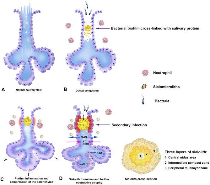
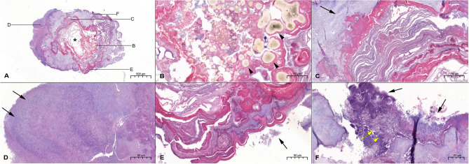
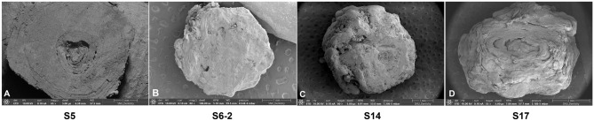
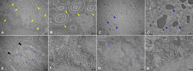
According to the previous studies of sialolithiasis reported so far, this study is aimed to identify the biological components of sialolith, which show different ultrastructures and chemical compositions from other stones, cholelith and urolith. Twenty-two specimens obtained from 20 patients were examined histologically, and analyzed with micro-CT, scanning electron microscopy (SEM), energy dispersive X-ray spectroscopy (EDS), and transmission electron microscopy (TEM). All sialoliths (n = 22) observed in this study showed a central nidus, which was filled with organoid matrix admixed with exosome vesicles, loose calcium apatite crystals, and many bacteria. The micro-CT and SEM observation clearly defined a single or multiple central nidus(es) encircled by highly calcified compact zone. The circular compact zone showed a band-like calcification, about 1-3 mm in thickness, and usually located between the central nidus and the peripheral multilayer zone. But some sialoliths (n = 5) showed severe erosion of compact zone by expanding multilayered zone depending on the level of calcification and inflammation in sialolith. By observing TEM images, many exosome vesicles and degraded cytoplasmic organelles were found in the central nidus, and some epithelial cells were also found in the calcified matrix of peripheral multilayer zone. Particularly, EDS analysis indicated the highest Ca/P ratio in the intermediate compact zone (1.77), and followed by the central nidus area (1.39) and the peripheral multilayer zone (0.87). Taken together, these data suggest that the central nidus containing many inflammatory exosomes and degraded cytoplasmic organelles has a potential to induce a band-like calcification of compact zone, and followed by the additional multilayer deposition of exfoliated salivary epithelial cells as well as salivary materials. Thereby, the calcium apatite-based sialolith is gradually growing in its volume size, and eventually obstructs the salivary flow and provides a site for the bacterial infection.
根据迄今为止报道的涎石病的先前研究,本研究旨在确定涎石的生物成分,这些成分的超微结构和化学成分与其他结石、胆石和尿石不同。从 20 名患者中获得的 22 个标本进行了组织学检查,并通过微计算机断层扫描(micro-CT)、扫描电子显微镜(SEM)、能谱分析(EDS)和透射电子显微镜(TEM)进行了分析。本研究中观察到的所有涎石(n=22)均显示中央核心,该核心充满了混合有外泌体囊泡的器官样基质、疏松的钙磷灰石晶体和许多细菌。micro-CT 和 SEM 观察清楚地定义了单个或多个中央核心(多个),被高度钙化的致密区环绕。圆形致密区显示出带状钙化,厚度约为 1-3mm,通常位于中央核心和周围多层区之间。但一些涎石(n=5)由于涎石中钙化和炎症的程度不同,显示出严重的致密区侵蚀,多层区扩展。通过观察 TEM 图像,在中央核心中发现了许多外泌体囊泡和降解的细胞质细胞器,在周围多层区的钙化基质中也发现了一些上皮细胞。特别是,EDS 分析表明中间致密区的 Ca/P 比值最高(1.77),其次是中央核心区(1.39)和周围多层区(0.87)。综上所述,这些数据表明,富含许多炎症性外泌体和降解的细胞质细胞器的中央核心具有诱导致密区带状钙化的潜力,并随后引发脱落的唾液上皮细胞以及唾液物质的多层沉积。因此,基于钙磷灰石的涎石逐渐增大其体积大小,并最终阻塞唾液流动并为细菌感染提供场所。