Jones Sarah, VandenHeuvel Sabrina, Luengo Martinez Andres, Birur Ruchi, Burgeson Eric, Gilbert Isabelle, Baker Aaron, Wolf Matthew, Raghavan Shreya A, Rogers Simon, Cosgriff-Hernandez Elizabeth
Department of Biomedical Engineering, The University of Texas at Austin, Austin, TX, 78712, USA.
Department of Biomedical Engineering, Texas A&M University, College Station, TX, 77843, USA.
Bioact Mater. 2024 Aug 28;41:640-656. doi: 10.1016/j.bioactmat.2024.08.012. eCollection 2024 Nov.
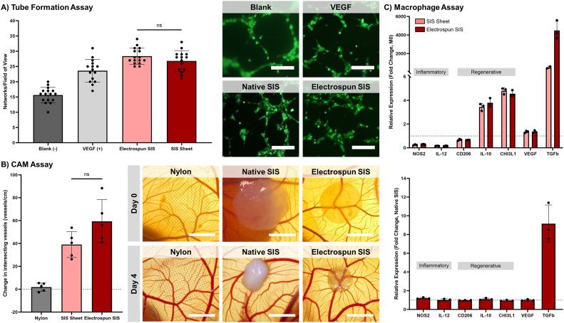
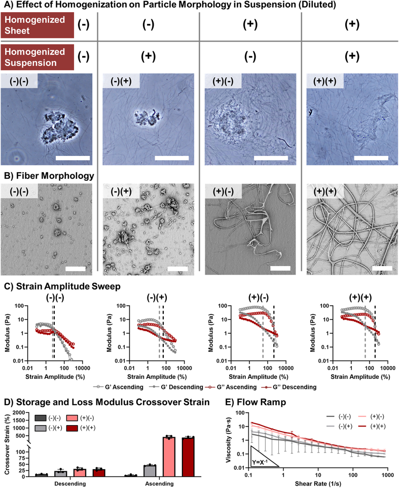
Decellularized extracellular matrices (dECM) have strong regenerative potential as tissue engineering scaffolds; however, current clinical options for dECM scaffolds are limited to freeze-drying its native form into sheets. Electrospinning is a versatile scaffold fabrication technique that allows control of macro- and microarchitecture. It remains challenging to electrospin dECM, which has led researchers to either blend it with synthetic materials or use enzymatic digestion to fully solubilize the dECM. Both strategies reduce the innate bioactivity of dECM and limit its regenerative potential. Herein, we developed a new suspension electrospinning method to fabricate a pure dECM fibrous mesh that retains its innate bioactivity. Systematic investigation of suspension parameters was used to identify critical rheological properties required to instill "spinnability," including homogenization, concentration, and particle size. Homogenization enhanced particle interaction to impart the requisite elastic behavior to withstand electrostatic drawing without breaking. A direct correlation between concentration and viscosity was observed that altered fiber morphology; whereas, particle size had minimal impact on suspension properties and fiber morphology. The versatility of this new method was demonstrated by electrospinning dECM with three common decellularization techniques (Abraham, Badylak, Luo) and tissue sources (intestinal submucosa, heart, skin). Bioactivity retention after electrospinning was confirmed using cell proliferation, angiogenesis, and macrophage polarization assays. Collectively, these findings provide a framework for researchers to electrospin dECM for diverse tissue engineering applications.
去细胞细胞外基质(dECM)作为组织工程支架具有强大的再生潜力;然而,目前dECM支架的临床选择仅限于将其天然形式冷冻干燥成片。静电纺丝是一种通用的支架制造技术,能够控制宏观和微观结构。对dECM进行静电纺丝仍然具有挑战性,这导致研究人员要么将其与合成材料混合,要么使用酶消化来使dECM完全溶解。这两种策略都会降低dECM的固有生物活性并限制其再生潜力。在此,我们开发了一种新的悬浮静电纺丝方法来制造保留其固有生物活性的纯dECM纤维网。通过对悬浮参数进行系统研究,以确定赋予“可纺性”所需的关键流变学特性,包括均匀化、浓度和粒径。均匀化增强了颗粒间的相互作用,赋予必要的弹性行为以承受静电拉伸而不破裂。观察到浓度与粘度之间存在直接相关性,这会改变纤维形态;而粒径对悬浮性能和纤维形态的影响最小。通过使用三种常见的去细胞技术(亚伯拉罕法、巴迪拉克法、罗法)和组织来源(肠黏膜下层、心脏、皮肤)对dECM进行静电纺丝,证明了这种新方法的通用性。使用细胞增殖、血管生成和巨噬细胞极化测定法证实了静电纺丝后生物活性的保留。总的来说,这些发现为研究人员将dECM静电纺丝用于各种组织工程应用提供了一个框架。