Department of Ophthalmology, Renmin Hospital of Wuhan University, Wuhan, China.
The First People's Hospital of Guangshui City, Guangshui, China.
Transl Vis Sci Technol. 2024 Sep 3;13(9):21. doi: 10.1167/tvst.13.9.21.
Using swept-source optical coherence tomography (SS-OCT) to explore the effect of high myopia on superficial retina vascular density (SVD) of the peripheral region and the area of radial peripapillary capillaries (RPCs).
In this cross-sectional study, a total of 91 volunteers (34 male subjects and 57 female subjects) were recruited and 34 individuals in the high myopic group (group A) and 57 individuals in the low myopic group (group B). Using the wide-field OCT-angiography (OCTA; 24 × 20 mm, 120 degrees angular field) compared the peripheral SVD and the area of RPC between the two groups and investigated its correlation with ocular axial length and diopter.
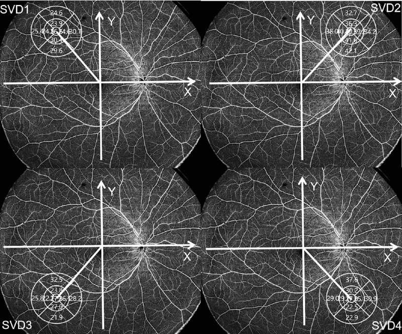
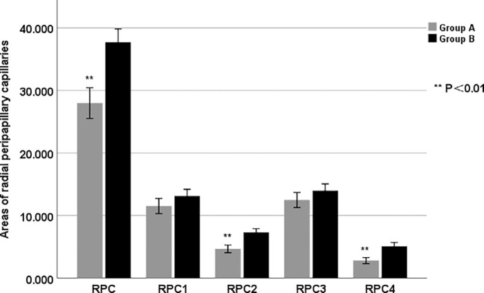
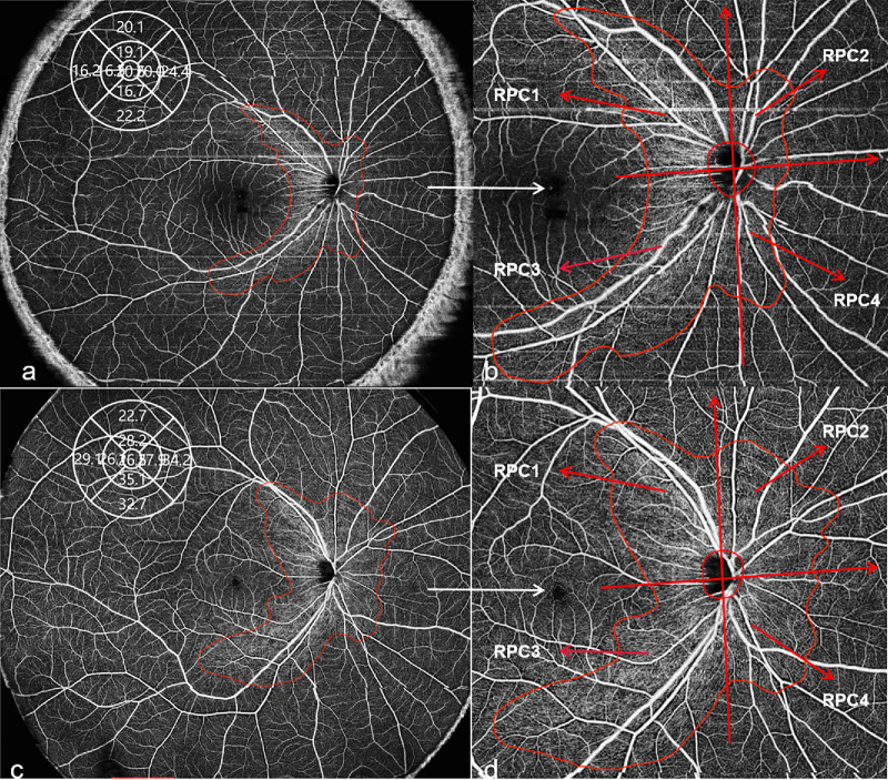
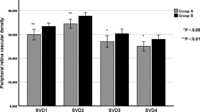
Peripheral SVD of group B around the supratemporal (SVD1), supranasal (SVD2), infratemporal (SVD3), and infranasal (SVD4) directions were significantly higher than those of group A (all P < 0.05). The RPC area of group B around the supranasal (RPC2) and infranasal (RPC4) were significantly larger than that of group A (all P < 0.01). Ocular axial length and diopter were significantly correlated with SVD2 and SVD4 (all P < 0.05), and they also have a significant correlation with the supratemporal (RPC1), RPC2, and RPC4 (all P < 0.05).
Peripheral SVD was decreased and the RPC area was mainly reduced on the nasal side in the high myopic group. Peripheral SVD and area of RPC are significantly correlated with ocular axial length and diopter.
The wide-field OCTA can be used for new detection of myopia's impact on the retinal peripheral SVD and area of peripapillary RPC, offering new insights into the progression of myopia.
利用扫频源光学相干断层扫描(SS-OCT)探讨高度近视对周边视网膜浅层血管密度(SVD)和视盘周围毛细血管(RPC)面积的影响。
本横断面研究共招募了 91 名志愿者(34 名男性和 57 名女性),其中 34 名高度近视患者(A 组)和 57 名低度近视患者(B 组)。使用广角 OCT 血管造影(OCTA;24×20mm,120°角视野)比较两组的周边 SVD 和 RPC 面积,并研究其与眼轴长度和屈光度的相关性。
B 组颞上(SVD1)、颞下(SVD3)、鼻上(SVD2)和鼻下(SVD4)方向的周边 SVD 明显高于 A 组(均 P<0.05)。B 组鼻上(RPC2)和鼻下(RPC4)的 RPC 面积明显大于 A 组(均 P<0.01)。眼轴长度和屈光度与 SVD2 和 SVD4 显著相关(均 P<0.05),与颞上(RPC1)、RPC2 和 RPC4 也有显著相关性(均 P<0.05)。
高度近视组周边 SVD 降低,鼻侧 RPC 面积主要减少。周边 SVD 和 RPC 面积与眼轴长度和屈光度显著相关。
崔秀华