Zhang Weiran, Li Chang, Gong Yibo, Liu Nianen, Cao Yunshan, Li Zhiqing, Zhang Yan
Tianjin Key Laboratory of Retinal Functions and Diseases, Tianjin Branch of National Clinical Research Center for Ocular Disease, Eye Institute and School of Optometry, Tianjin Medical University Eye Hospital, Tianjin, China.
Department of Cardiology, Gansu Provincial Hospital, Lanzhou, China.
Front Bioeng Biotechnol. 2022 Aug 16;10:920197. doi: 10.3389/fbioe.2022.920197. eCollection 2022.
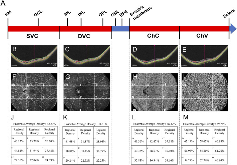
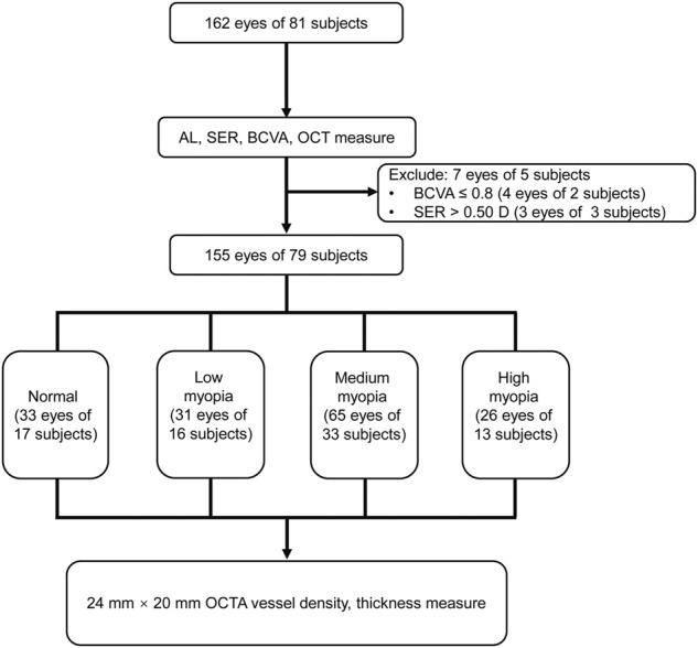
To detect previously undetectable changes in vessel density and structural thickness, the two biomechanics-related parameters reflecting hemodynamics and tensile strength, respectively, in the peripheral and central fundi of nonpathological myopic eyes with an advanced ultrawide-field optical coherence tomography angiography (OCTA) system. A cross-sectional observational clinical study was carried out by recruiting 155 eyes from 79 college students aged 18-28 years. The eyes were stratified into normal, low-myopia, medium-myopia, and high-myopia groups according to diopter. A newly developed OCTA system with scanning dimensions of 24 mm × 20 mm, acquisition speed of 400 kHz, and imaging range of 6 mm was used to examine the vessel densities of superficial vascular complex (SVC), deep vascular complex (DVC), choriocapillary (ChC), and choroidal vessel (ChV) layers, as well as the thicknesses of the inner retina, outer retina, and choroid in the nonpathological myopic eyes. The vessel densities in ChV at the temporal, inferotemporal, inferior, and inferonasal regions in the fundus periphery were significantly reduced in myopic subjects as compared to normal controls (all < 0.05). The thicknesses of the inner retinal segments in most peripheral regions of the fundus became attenuated along with myopia severity (all < 0.05). The thicknesses of the outer retinal segments were diminished at the superior and supranasal regions of the peripheral fundi of myopic subjects as compared to normal controls (all < 0.05). At the central macular region, the decreased vessel densities of SVC and DVC were correlated with the attenuated thicknesses of inner retinal segments, respectively (all < 0.05). As revealed for the first time by the advanced ultrawide-field OCTA system, the two biomechanics-related parameters that include the densities of the choroidal vessels and thicknesses of the inner retina segments were significantly reduced in the periphery of nonpathological myopic fundi and the reductions were associated with myopia severity. At the central macular region, the newly developed device provides consistent results with the previous findings. Therefore, it is important to use the noninvasive, ultrawide-field OCTA with high resolution for early detection of fundus changes in subjects with nonpathological high myopia. clinicaltrials.gov, identifier ChiCTR2100054093.
为了检测血管密度和结构厚度中先前无法检测到的变化,这两个生物力学相关参数分别反映了非病理性近视眼中周边和中央眼底的血流动力学和拉伸强度,采用了先进的超广角光学相干断层扫描血管造影(OCTA)系统。通过招募79名年龄在18 - 28岁的大学生的155只眼睛进行了一项横断面观察性临床研究。根据屈光度将眼睛分为正常、低度近视、中度近视和高度近视组。使用一种新开发的OCTA系统,其扫描尺寸为24 mm×20 mm,采集速度为400 kHz,成像范围为6 mm,来检查非病理性近视眼中浅表血管复合体(SVC)、深层血管复合体(DVC)、脉络膜毛细血管(ChC)和脉络膜血管(ChV)层的血管密度,以及视网膜内层、外层和脉络膜的厚度。与正常对照组相比,近视受试者眼底周边颞侧、颞下、下方和鼻下区域的ChV血管密度显著降低(均P<0.05)。随着近视严重程度的增加,眼底大多数周边区域的视网膜内层厚度变薄(均P<0.05)。与正常对照组相比,近视受试者周边眼底上方和鼻上区域的视网膜外层厚度减小(均P<0.05)。在中央黄斑区,SVC和DVC血管密度的降低分别与视网膜内层厚度的变薄相关(均P<0.05)。先进的超广角OCTA系统首次揭示,在非病理性近视眼底周边,包括脉络膜血管密度和视网膜内层厚度在内的两个生物力学相关参数显著降低,且这些降低与近视严重程度相关。在中央黄斑区,新开发的设备提供了与先前研究结果一致的结果。因此,使用具有高分辨率的无创超广角OCTA对于早期检测非病理性高度近视患者的眼底变化很重要。ClinicalTrials.gov标识符:ChiCTR2100054093。