School of Biotechnology, Kalinga Institute of Industrial Technology, Deemed to Be University, Bhubaneswar, Odisha, 751024, India.
Department of Pediatrics, Kalinga Institute of Medical Sciences, Deemed to Be University, Bhubaneswar, Odisha, 751024, India.
Cell Commun Signal. 2024 Sep 26;22(1):451. doi: 10.1186/s12964-024-01779-4.
Dengue is a vector-borne debilitating disease that is manifested as mild dengue fever, dengue with warning signs, and severe dengue. Dengue infection provokes a collective immune response; in particular, the innate immune response plays a key role in primary infection and adaptive immunity during secondary infection. In this review, we comprehensively walk through the various markers of immune response against dengue pathogenesis and outcome.
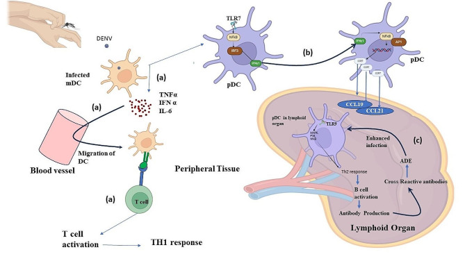
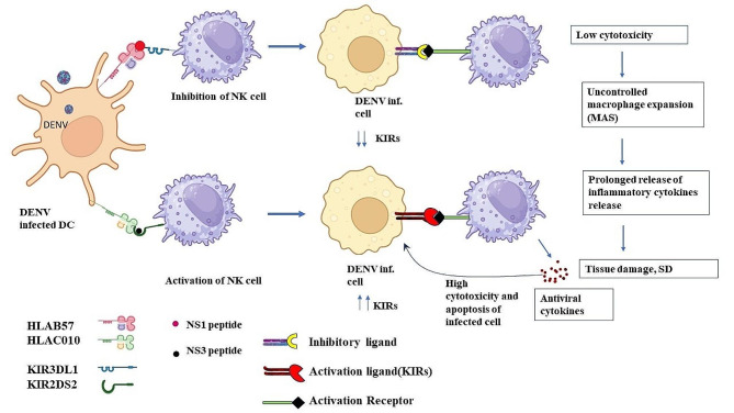
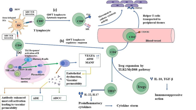
Innate immune response against dengue involves a collective response through the expression of proinflammatory cytokines, such as tumor necrosis factors (TNFs), interferons (IFNs), and interleukins (ILs), in addition to anti-inflammatory cytokines and toll-like receptors (TLRs) in modulating viral pathogenesis. Monocytes, dendritic cells (DCs), and mast cells are the primary innate immune cells initially infected by DENV. Such immune cells modulate the expression of various markers, which can influence disease severity by aiding virus entry and proinflammatory responses. Adaptive immune response is mainly aided by B and T lymphocytes, which stimulate the formation of germinal centers for plasmablast development and antibody production. Such antibodies are serotype-dependent and can aid in virus entry during secondary infection, mediated through a different serotype, such as in antibody-dependent enhancement (ADE), leading to DENV severity. The entire immunological repertoire is exhibited differently depending on the immune status of the individual.
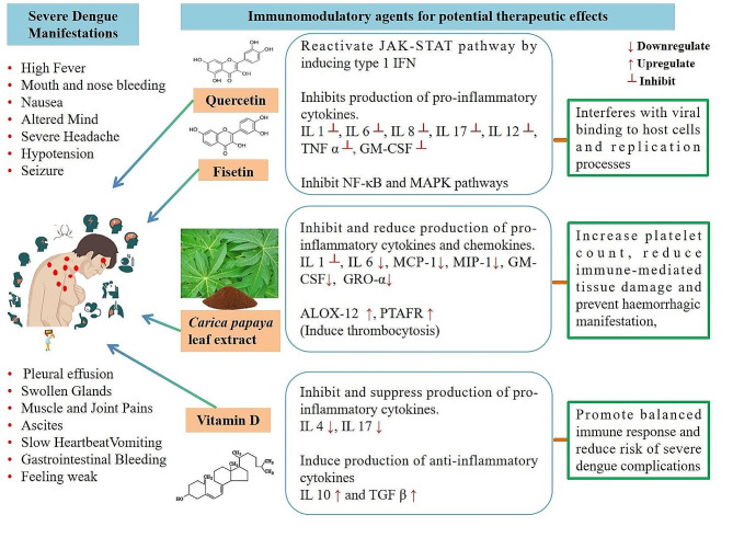
Dengue fever through severe dengue proceeds along with the modulated expression of several immune markers. In particular, TLR2, TNF-α, IFN-I, IL-6, IL-8, IL-17 and IL-10, in addition to intermediate monocytes (CD14+CD16+) and Th17 (CD4+IL-17+) cells are highly expressed during severe dengue. Such markers could assist greatly in severity assessment, prompt diagnosis, and treatment.
登革热是一种由蚊子传播的使人虚弱的疾病,表现为轻度登革热、有警告信号的登革热和重症登革热。登革热感染会引发集体免疫反应;特别是,固有免疫反应在初次感染和二次感染时的适应性免疫中发挥关键作用。在这篇综述中,我们全面了解了针对登革热发病机制和结果的各种免疫反应标志物。
针对登革热的固有免疫反应涉及通过表达促炎细胞因子(如肿瘤坏死因子[TNFs]、干扰素[IFNs]和白细胞介素[ILs])的集体反应,此外还涉及调节病毒发病机制的抗炎细胞因子和 toll 样受体(TLRs)。单核细胞、树突状细胞(DCs)和肥大细胞是最初感染 DENV 的主要固有免疫细胞。这些免疫细胞调节各种标志物的表达,通过帮助病毒进入和促炎反应来影响疾病严重程度。适应性免疫反应主要由 B 和 T 淋巴细胞辅助,它们刺激生发中心的形成,促进浆母细胞的发育和抗体的产生。这些抗体是血清型依赖性的,可以在二次感染期间通过不同的血清型(如抗体依赖性增强[ADE])帮助病毒进入,导致登革热的严重程度。整个免疫谱根据个体的免疫状态表现不同。
登革热从轻症到重症的发展伴随着几种免疫标志物的调节表达。特别是 TLR2、TNF-α、IFN-I、IL-6、IL-8、IL-17 和 IL-10,以及中间单核细胞(CD14+CD16+)和 Th17(CD4+IL-17+)细胞在重症登革热中高度表达。这些标志物可以极大地帮助进行严重程度评估、及时诊断和治疗。