• 文献检索
  • 文档翻译
  • 深度研究
  • 学术资讯
  • Suppr Zotero 插件Zotero 插件
  • 邀请有礼
  • 套餐&价格
  • 历史记录
应用&插件
Suppr Zotero 插件Zotero 插件浏览器插件Mac 客户端Windows 客户端微信小程序
定价
高级版会员购买积分包购买API积分包
服务
文献检索文档翻译深度研究API 文档MCP 服务
关于我们
关于 Suppr公司介绍联系我们用户协议隐私条款
关注我们

Suppr 超能文献

核心技术专利:CN118964589B侵权必究
粤ICP备2023148730 号-1Suppr @ 2026

文献检索

告别复杂PubMed语法,用中文像聊天一样搜索,搜遍4000万医学文献。AI智能推荐,让科研检索更轻松。

立即免费搜索

文件翻译

保留排版,准确专业,支持PDF/Word/PPT等文件格式,支持 12+语言互译。

免费翻译文档

深度研究

AI帮你快速写综述,25分钟生成高质量综述,智能提取关键信息,辅助科研写作。

立即免费体验

视网膜外层带的偏振分辨分析:使用全场扫频源光学相干断层扫描研究弹道光子和多次散射光子

Polarization-resolved analysis of outer retinal bands: investigating ballistic and multiply scattered photons using full-field swept-source optical coherence tomography.

作者信息

Ahmed Shaiban, Son Taeyoon, Yao Xincheng

机构信息

Department of Biomedical Engineering, University of Illinois Chicago, Chicago, IL 60607, USA.

Department of Ophthalmology and Visual Sciences, University of Illinois Chicago, Chicago, IL 60612, USA.

出版信息

Biomed Opt Express. 2024 Jul 18;15(8):4749-4763. doi: 10.1364/BOE.523202. eCollection 2024 Aug 1.

DOI:10.1364/BOE.523202
PMID:39346986
原文链接:https://pmc.ncbi.nlm.nih.gov/articles/PMC11427207/
Abstract

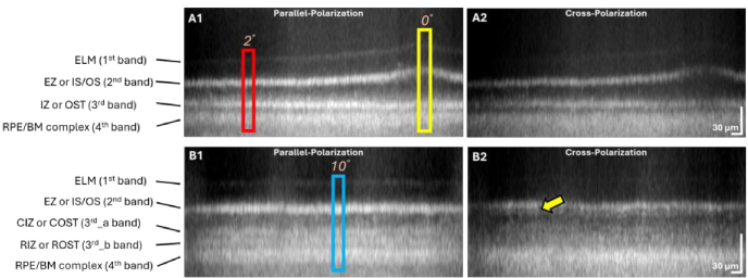
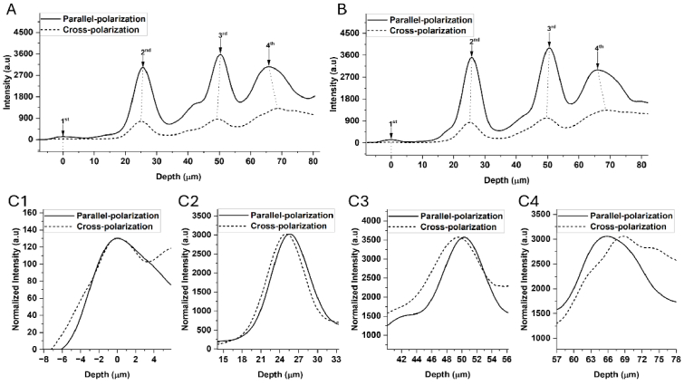
Precise interpretation of the anatomical origins of outer retinal optical coherence tomography (OCT) presents technical challenges owing to the delicate nature of the retina. To address this challenge, our study introduces a novel polarization-sensitive full-field swept-source OCT (FF-SS-OCT) that provides parallel-polarization and cross-polarization OCT measurements, predominantly capturing ballistically reflected photons and multiply scattered photons, respectively. Notably, parallel-polarization OCT unveils layer-like structures more effectively, including the inner plexiform layer (IPL) sub-layers, outer plexiform layer (OPL) sub-layers (in rod-dominant regions), and rod/cone outer segment (OS) tips, compared to cross-polarization OCT, where such sub-layers are not visible. Through a comparative analysis of parallel-polarization and cross-polarization OCT images of the outer retina, we discovered that the 2 outer retinal OCT band results from contributions from both the ellipsoid zone (EZ) and the inner segment/outer segment (IS/OS) junction. Similarly, the 3 outer retinal OCT band appears to reflect contributions from both the interdigitation zone (IZ) and photoreceptor OS tips. This polarization-sensitive approach advances our understanding of the origins of outer retinal OCT signals and proposes potential new biomarkers for assessing retinal health and diseases.

摘要

由于视网膜的精细特性,对外侧视网膜光学相干断层扫描(OCT)的解剖学起源进行精确解读存在技术挑战。为应对这一挑战,我们的研究引入了一种新型偏振敏感全场扫频源OCT(FF-SS-OCT),它可提供平行偏振和交叉偏振OCT测量,分别主要捕获弹道反射光子和多次散射光子。值得注意的是,与交叉偏振OCT相比,平行偏振OCT能更有效地揭示层状结构,包括内网状层(IPL)子层、外网状层(OPL)子层(在视杆细胞占主导的区域)以及视杆/视锥细胞外段(OS)尖端,而在交叉偏振OCT中这些子层不可见。通过对外侧视网膜平行偏振和交叉偏振OCT图像的对比分析,我们发现外侧视网膜OCT的两条带是由椭圆体带(EZ)和内段/外段(IS/OS)交界处共同作用产生的。同样,外侧视网膜OCT的三条带似乎反映了指状交叉区(IZ)和光感受器OS尖端的共同作用。这种偏振敏感方法增进了我们对外侧视网膜OCT信号起源的理解,并提出了用于评估视网膜健康和疾病的潜在新生物标志物。

https://cdn.ncbi.nlm.nih.gov/pmc/blobs/c0e9/11427207/1842cfd8009b/boe-15-8-4749-g007.jpg
https://cdn.ncbi.nlm.nih.gov/pmc/blobs/c0e9/11427207/ded5ca873a21/boe-15-8-4749-g001.jpg
https://cdn.ncbi.nlm.nih.gov/pmc/blobs/c0e9/11427207/93ce25439a2b/boe-15-8-4749-g002.jpg
https://cdn.ncbi.nlm.nih.gov/pmc/blobs/c0e9/11427207/8e9b422234fe/boe-15-8-4749-g003.jpg
https://cdn.ncbi.nlm.nih.gov/pmc/blobs/c0e9/11427207/c22d13b68954/boe-15-8-4749-g004.jpg
https://cdn.ncbi.nlm.nih.gov/pmc/blobs/c0e9/11427207/516ead66ce12/boe-15-8-4749-g005.jpg
https://cdn.ncbi.nlm.nih.gov/pmc/blobs/c0e9/11427207/bdc698cd7157/boe-15-8-4749-g006.jpg
https://cdn.ncbi.nlm.nih.gov/pmc/blobs/c0e9/11427207/1842cfd8009b/boe-15-8-4749-g007.jpg

相似文献

1
Polarization-resolved analysis of outer retinal bands: investigating ballistic and multiply scattered photons using full-field swept-source optical coherence tomography.视网膜外层带的偏振分辨分析:使用全场扫频源光学相干断层扫描研究弹道光子和多次散射光子
Biomed Opt Express. 2024 Jul 18;15(8):4749-4763. doi: 10.1364/BOE.523202. eCollection 2024 Aug 1.
2
Ganglion Cell Layer Thickness as a Biomarker for Amyotrophic Lateral Sclerosis Functional Outcome: An OCT study.神经节细胞层厚度作为肌萎缩侧索硬化症功能预后的生物标志物:一项光学相干断层扫描研究。
Rom J Ophthalmol. 2025 Apr-Jun;69(2):200-207. doi: 10.22336/rjo.2025.32.
3
Polarization optical coherence tomography optoretinography: verifying light-induced photoreceptor outer segment shrinkage and subretinal space expansion.偏振光学相干断层扫描视网膜电图:验证光诱导的光感受器外段收缩和视网膜下间隙扩大。
Neurophotonics. 2025 Jan;12(1):015005. doi: 10.1117/1.NPh.12.1.015005. Epub 2025 Jan 27.
4
Visualizing the invisible: inner plexiform layer stratification with conventional spectral-domain optical coherence tomography.可视化不可见之物:利用传统光谱域光学相干断层扫描技术对内核层进行分层
Int J Retina Vitreous. 2025 Jun 15;11(1):65. doi: 10.1186/s40942-025-00692-3.
5
Optical coherence tomography (OCT) for detection of macular oedema in patients with diabetic retinopathy.光学相干断层扫描(OCT)用于检测糖尿病视网膜病变患者的黄斑水肿。
Cochrane Database Syst Rev. 2015 Jan 7;1(1):CD008081. doi: 10.1002/14651858.CD008081.pub3.
6
Early inner plexiform layer thinning and retinal nerve fiber layer thickening in excitotoxic retinal injury using deep learning-assisted optical coherence tomography.深度学习辅助光学相干断层扫描在兴奋性视网膜损伤中的早期内丛状层变薄和视网膜神经纤维层增厚。
Acta Neuropathol Commun. 2024 Feb 1;12(1):19. doi: 10.1186/s40478-024-01732-z.
7
Inner Plexiform Layer Substrata Are Discernible with Commercial OCT and Affected by Aging.内网状层基质可通过商用光学相干断层扫描(OCT)识别且受衰老影响。
Ophthalmol Sci. 2025 Apr 28;5(5):100815. doi: 10.1016/j.xops.2025.100815. eCollection 2025 Sep-Oct.
8
Adaptive spectroscopic visible-light optical coherence tomography for clinical retinal oximetry.用于临床视网膜血氧测定的自适应光谱可见光光学相干断层扫描技术。
Commun Med (Lond). 2023 Apr 24;3(1):57. doi: 10.1038/s43856-023-00288-8.
9
Retinal layer segmentation in multiple sclerosis: a systematic review and meta-analysis.多发性硬化症中的视网膜层分割:系统评价和荟萃分析。
Lancet Neurol. 2017 Oct;16(10):797-812. doi: 10.1016/S1474-4422(17)30278-8. Epub 2017 Sep 12.
10
Longitudinal Assessment of Ocular Biomarkers in Individuals With Gulf War Illness Symptoms.海湾战争疾病症状个体眼部生物标志物的纵向评估。
Mil Med. 2024 Oct 3. doi: 10.1093/milmed/usae457.

引用本文的文献

1
Characterization of the depolarization contrast line in the outer retina using polarization-sensitive optical coherence tomography.使用偏振敏感光学相干断层扫描技术对外侧视网膜中的去极化对比线进行表征。
Sci Rep. 2025 Jul 14;15(1):25394. doi: 10.1038/s41598-025-11111-w.
2
Polarization optical coherence tomography optoretinography: verifying light-induced photoreceptor outer segment shrinkage and subretinal space expansion.偏振光学相干断层扫描视网膜电图:验证光诱导的光感受器外段收缩和视网膜下间隙扩大。
Neurophotonics. 2025 Jan;12(1):015005. doi: 10.1117/1.NPh.12.1.015005. Epub 2025 Jan 27.

本文引用的文献

1
Preserving polarization maintaining photons for enhanced contrast imaging of the retina.保留偏振态以维持光子用于增强视网膜的对比度成像。
Biomed Opt Express. 2023 Oct 24;14(11):5932-5945. doi: 10.1364/BOE.501636. eCollection 2023 Nov 1.
2
Spatially offset optical coherence tomography: Leveraging multiple scattering for high-contrast imaging at depth in turbid media.空间偏移光学相干断层扫描:利用多重散射在混浊介质中进行深度高对比度成像。
Sci Adv. 2023 Jul 7;9(27):eadh5435. doi: 10.1126/sciadv.adh5435.
3
Spatio-temporal optical coherence tomography provides full thickness imaging of the chorioretinal complex.
时空光学相干断层扫描提供脉络膜视网膜复合体的全层成像。
iScience. 2022 Nov 5;25(12):105513. doi: 10.1016/j.isci.2022.105513. eCollection 2022 Dec 22.
4
Visible Light Optical Coherence Tomography Reveals the Relationship of the Myoid and Ellipsoid to Band 2 in Humans.可见光是相干断层扫描揭示人类肌样体和椭圆体与带 2 的关系。
Transl Vis Sci Technol. 2022 Sep 1;11(9):3. doi: 10.1167/tvst.11.9.3.
5
Subcellular Comparison of Visible-Light Optical Coherence Tomography and Electron Microscopy in the Mouse Outer Retina.亚细胞水平可见 光 光 学 相干 断层 扫描 和电镜 在 小鼠 外 视网膜 的 比较。
Invest Ophthalmol Vis Sci. 2022 Aug 2;63(9):10. doi: 10.1167/iovs.63.9.10.
6
Methods and applications of full-field optical coherence tomography: a review.全场光学相干断层扫描方法与应用:综述。
J Biomed Opt. 2022 May;27(5). doi: 10.1117/1.JBO.27.5.050901.
7
ADC-Net: An Open-Source Deep Learning Network for Automated Dispersion Compensation in Optical Coherence Tomography.ADC-Net:一种用于光学相干断层扫描中自动色散补偿的开源深度学习网络。
Front Med (Lausanne). 2022 Apr 8;9:864879. doi: 10.3389/fmed.2022.864879. eCollection 2022.
8
Adaptive optics for high-resolution imaging.用于高分辨率成像的自适应光学技术。
Nat Rev Methods Primers. 2021;1. doi: 10.1038/s43586-021-00066-7. Epub 2021 Oct 14.
9
The 3D organisation of mitochondria in primate photoreceptors.灵长类光感受器中线粒体的 3D 组织。
Sci Rep. 2021 Sep 22;11(1):18863. doi: 10.1038/s41598-021-98409-7.
10
Interpretation of anatomic correlates of outer retinal bands in optical coherence tomography.光学相干断层扫描中外层视网膜带的解剖学相关性解读。
Exp Biol Med (Maywood). 2021 Oct;246(20):2140-2150. doi: 10.1177/15353702211022674. Epub 2021 Jun 10.