Guerra Ricardo Luz Leitão, Roisman Luiz, Duker Jay S, Querques Giuseppe, Lucatto Luiz Filipe Adami, Badaró Emmerson, Barbosa Gabriel Castilho S, Novais Eduardo Amorim
Retina Department, Leitão Guerra - Oftalmologia, Salvador, Brazil.
Orbit Ophthalmo Learning, Salvador, Brazil.
Int J Retina Vitreous. 2025 Jun 15;11(1):65. doi: 10.1186/s40942-025-00692-3.
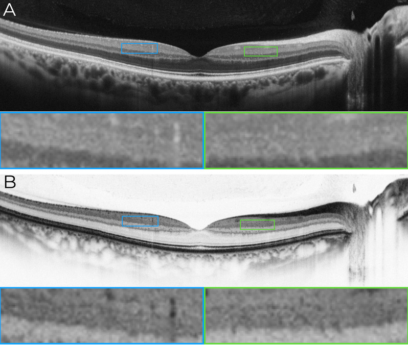
The inner plexiform layer (IPL) of the retina plays a key role in visual processing, consisting of five stratified sub-bands (S1-S5) that segregate ON and OFF visual pathways. Until now, resolving these IPL sub-layers was only possible with experimental high-resolution (HR-OCT) or visible-light OCT (VIS-OCT), which remain inaccessible for clinical use. This study provides the first demonstration that IPL stratification can be visualized using commercially available spectral-domain OCT (SD-OCT) with optimized imaging and grayscale inversion.
This retrospective, cross-sectional image analysis study included three healthy individuals who underwent macular OCT imaging. Two subjects were imaged with SD-OCT devices (Nidek RS3000 Advance and Zeiss Cirrus 6000), while one subject was imaged with a swept-source OCT (SS-OCT) device (Topcon Triton DRI). High-density B-scans (1024 A-scans per B-scan) with 120 repetitions for noise reduction were analyzed in both standard and inverted grayscale display modes. The impact of scan size (12 mm, 6 mm, and 3 mm) on IPL visualization was also evaluated.
In conventional grayscale, IPL stratification was indistinct. However, inverted grayscale revealed five IPL sub-bands in all cases, particularly in the parafoveal region where the IPL is thicker. Hyperreflective dots near IPL-1, likely representing the superficial capillary plexus, were also identified. The 3-mm scan protocol provided superior sub-layer differentiation compared to 12-mm scans. However, SS-OCT images did not allow for the distinction of the five IPL strata.
This study challenges the belief that IPL stratification cannot be identified with conventional SD-OCT. By refining imaging parameters and using grayscale inversion, this approach enhances retinal circuit analysis with standard technology. While SD-OCT enables detailed IPL visualization under specific conditions, SS-OCT does not appear to be well-suited for this purpose. These findings redefine SD-OCT's diagnostic capabilities, opening avenues for research in ophthalmology and neurodegenerative disease monitoring. Further studies should establish best practices and expand clinical applications for this novel methodology.
视网膜的内网状层(IPL)在视觉处理中起关键作用,由五个分层子带(S1 - S5)组成,这些子带分离开视觉通路和关视觉通路。到目前为止,只有通过实验性高分辨率光学相干断层扫描(HR - OCT)或可见光光学相干断层扫描(VIS - OCT)才能分辨这些IPL子层,而这些技术在临床应用中仍然无法实现。本研究首次证明,使用具有优化成像和灰度反转的商用光谱域光学相干断层扫描(SD - OCT)可以可视化IPL分层。
这项回顾性横断面图像分析研究纳入了三名接受黄斑OCT成像的健康个体。两名受试者使用SD - OCT设备(尼德克RS3000 Advance和蔡司Cirrus 6000)进行成像,而一名受试者使用扫频光学相干断层扫描(SS - OCT)设备(拓普康Triton DRI)进行成像。在标准和反转灰度显示模式下分析了具有120次重复以降低噪声的高密度B扫描(每次B扫描1024次A扫描)。还评估了扫描大小(12毫米、6毫米和3毫米)对IPL可视化的影响。
在传统灰度下,IPL分层不清晰。然而,反转灰度在所有情况下都显示出五个IPL子带,特别是在IPL较厚的黄斑旁区域。还识别出了IPL - 1附近的高反射点,可能代表浅表毛细血管丛。与12毫米扫描相比,3毫米扫描方案提供了更好的子层区分。然而,SS - OCT图像无法区分五个IPL层。
本研究挑战了传统SD - OCT无法识别IPL分层的观点。通过优化成像参数和使用灰度反转,这种方法增强了使用标准技术进行的视网膜回路分析。虽然SD - OCT在特定条件下能够实现详细的IPL可视化,但SS - OCT似乎不太适合此目的。这些发现重新定义了SD - OCT的诊断能力,为眼科研究和神经退行性疾病监测开辟了道路。进一步的研究应建立最佳实践并扩大这种新方法的临床应用。