• 文献检索
  • 文档翻译
  • 深度研究
  • 学术资讯
  • Suppr Zotero 插件Zotero 插件
  • 邀请有礼
  • 套餐&价格
  • 历史记录
应用&插件
Suppr Zotero 插件Zotero 插件浏览器插件Mac 客户端Windows 客户端微信小程序
定价
高级版会员购买积分包购买API积分包
服务
文献检索文档翻译深度研究API 文档MCP 服务
关于我们
关于 Suppr公司介绍联系我们用户协议隐私条款
关注我们

Suppr 超能文献

核心技术专利:CN118964589B侵权必究
粤ICP备2023148730 号-1Suppr @ 2026

文献检索

告别复杂PubMed语法,用中文像聊天一样搜索,搜遍4000万医学文献。AI智能推荐,让科研检索更轻松。

立即免费搜索

文件翻译

保留排版,准确专业,支持PDF/Word/PPT等文件格式,支持 12+语言互译。

免费翻译文档

深度研究

AI帮你快速写综述,25分钟生成高质量综述,智能提取关键信息,辅助科研写作。

立即免费体验

连接组重建可预测用于导航的视觉特征。

Connectomic reconstruction predicts visual features used for navigation.

作者信息

Garner Dustin, Kind Emil, Lai Jennifer Yuet Ha, Nern Aljoscha, Zhao Arthur, Houghton Lucy, Sancer Gizem, Wolff Tanya, Rubin Gerald M, Wernet Mathias F, Kim Sung Soo

机构信息

Molecular, Cellular, and Developmental Biology, University of California Santa Barbara, Santa Barbara, CA, USA.

Department of Biology, Freie Universität Berlin, Berlin, Germany.

出版信息

Nature. 2024 Oct;634(8032):181-190. doi: 10.1038/s41586-024-07967-z. Epub 2024 Oct 2.

DOI:10.1038/s41586-024-07967-z
PMID:39358517
原文链接:https://pmc.ncbi.nlm.nih.gov/articles/PMC11446847/
Abstract

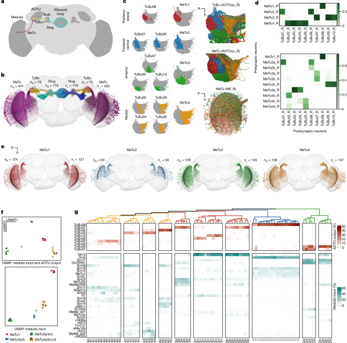
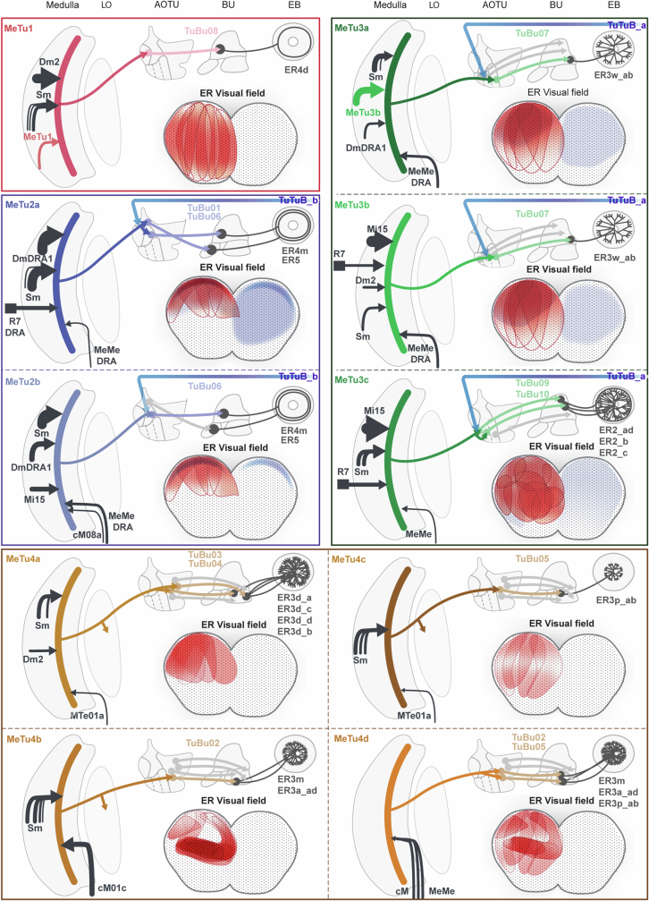
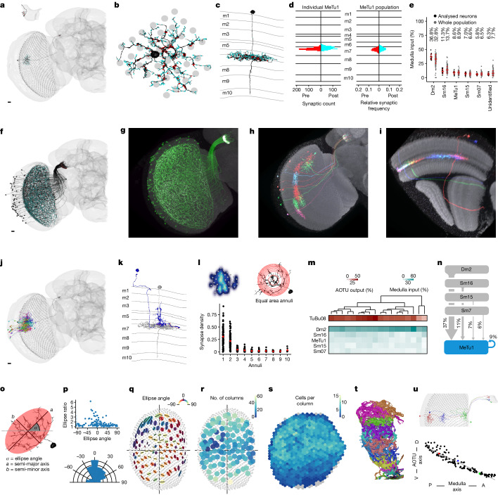
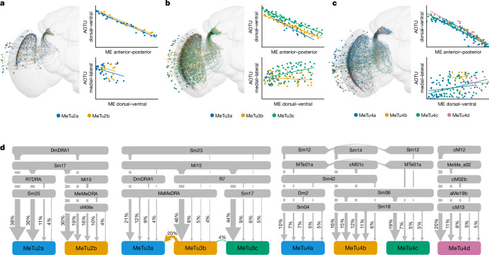
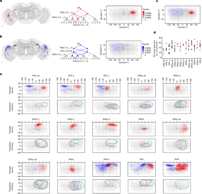
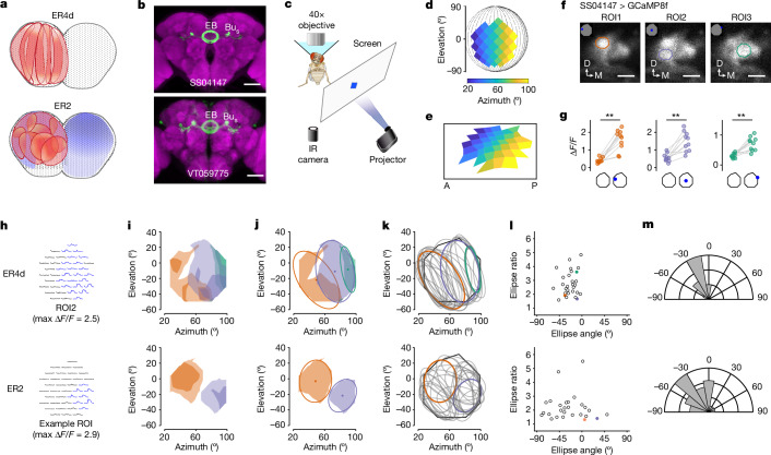
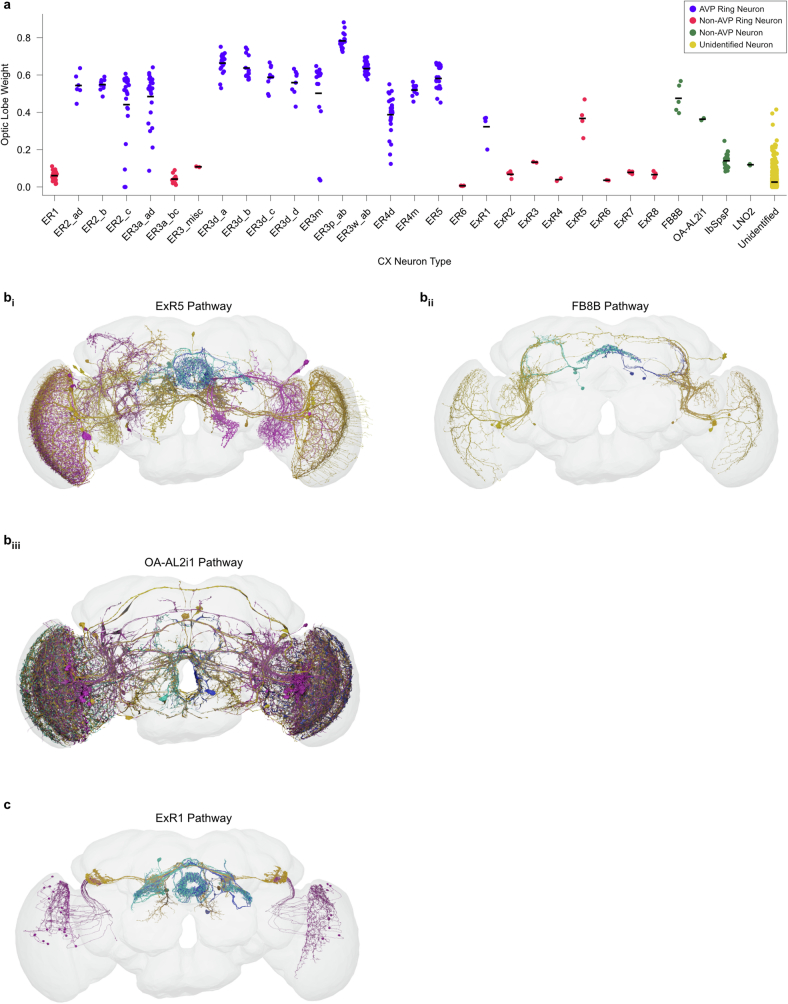
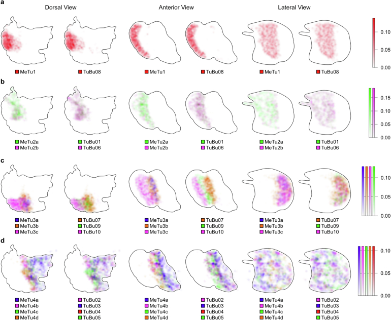
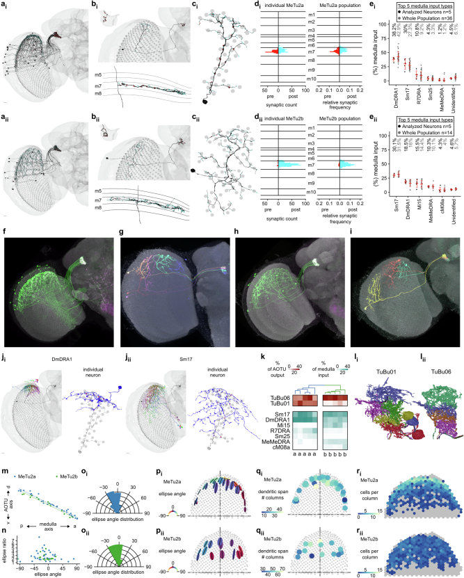
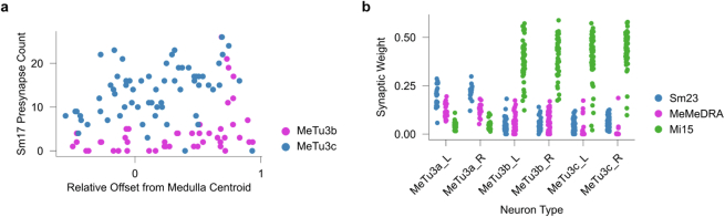
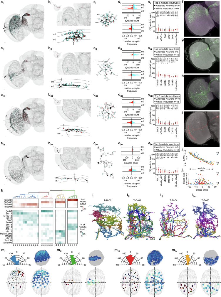
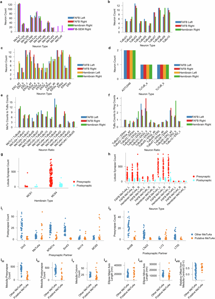
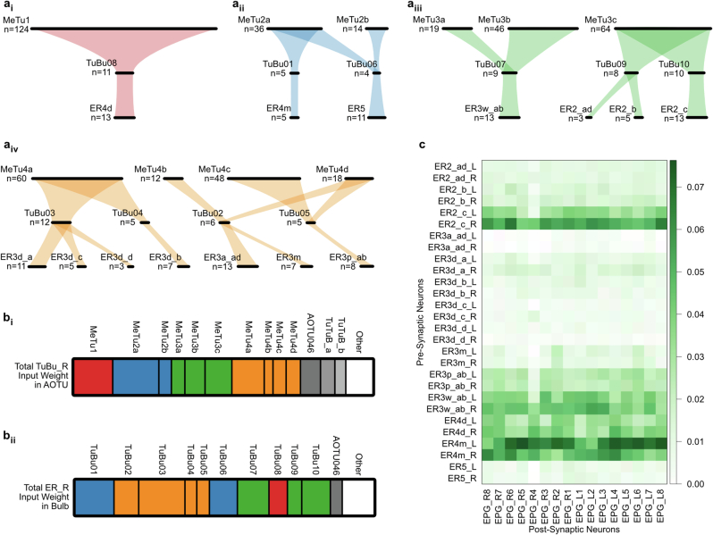
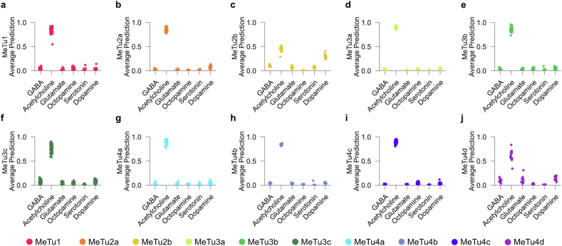
Many animals use visual information to navigate, but how such information is encoded and integrated by the navigation system remains incompletely understood. In Drosophila melanogaster, EPG neurons in the central complex compute the heading direction by integrating visual input from ER neurons, which are part of the anterior visual pathway (AVP). Here we densely reconstruct all neurons in the AVP using electron-microscopy data. The AVP comprises four neuropils, sequentially linked by three major classes of neurons: MeTu neurons, which connect the medulla in the optic lobe to the small unit of the anterior optic tubercle (AOTUsu) in the central brain; TuBu neurons, which connect the AOTUsu to the bulb neuropil; and ER neurons, which connect the bulb to the EPG neurons. On the basis of morphologies, connectivity between neural classes and the locations of synapses, we identify distinct information channels that originate from four types of MeTu neurons, and we further divide these into ten subtypes according to the presynaptic connections in the medulla and the postsynaptic connections in the AOTUsu. Using the connectivity of the entire AVP and the dendritic fields of the MeTu neurons in the optic lobes, we infer potential visual features and the visual area from which any ER neuron receives input. We confirm some of these predictions physiologically. These results provide a strong foundation for understanding how distinct sensory features can be extracted and transformed across multiple processing stages to construct higher-order cognitive representations.

摘要

许多动物利用视觉信息进行导航,但导航系统如何对这些信息进行编码和整合仍未完全了解。在黑腹果蝇中,中央复合体中的EPG神经元通过整合来自ER神经元的视觉输入来计算头部方向,ER神经元是前视觉通路(AVP)的一部分。在这里,我们利用电子显微镜数据对AVP中的所有神经元进行了密集重建。AVP由四个神经纤维网组成,通过三类主要神经元依次相连:MeTu神经元,连接视叶中的髓质与中脑前视结节小单元(AOTUsu);TuBu神经元,连接AOTUsu与球茎神经纤维网;以及ER神经元,连接球茎与EPG神经元。基于形态学、神经类之间的连接性以及突触的位置,我们识别出源自四种MeTu神经元的不同信息通道,并根据髓质中的突触前连接和AOTUsu中的突触后连接将这些通道进一步分为十个亚型。利用整个AVP的连接性以及视叶中MeTu神经元的树突场,我们推断出任何ER神经元接收输入的潜在视觉特征和视觉区域。我们通过生理学方法证实了其中一些预测。这些结果为理解如何在多个处理阶段提取和转换不同的感官特征以构建高阶认知表征提供了坚实的基础。

https://cdn.ncbi.nlm.nih.gov/pmc/blobs/2957/11446847/7ddee37f62ec/41586_2024_7967_Fig18_ESM.jpg
https://cdn.ncbi.nlm.nih.gov/pmc/blobs/2957/11446847/62c723f9642f/41586_2024_7967_Fig1_HTML.jpg
https://cdn.ncbi.nlm.nih.gov/pmc/blobs/2957/11446847/c45f997e9b7b/41586_2024_7967_Fig2_HTML.jpg
https://cdn.ncbi.nlm.nih.gov/pmc/blobs/2957/11446847/66db9092fbfc/41586_2024_7967_Fig3_HTML.jpg
https://cdn.ncbi.nlm.nih.gov/pmc/blobs/2957/11446847/a9161b89a0d6/41586_2024_7967_Fig4_HTML.jpg
https://cdn.ncbi.nlm.nih.gov/pmc/blobs/2957/11446847/4250a88757e9/41586_2024_7967_Fig5_HTML.jpg
https://cdn.ncbi.nlm.nih.gov/pmc/blobs/2957/11446847/daebd2307634/41586_2024_7967_Fig6_ESM.jpg
https://cdn.ncbi.nlm.nih.gov/pmc/blobs/2957/11446847/6a42ed277364/41586_2024_7967_Fig7_ESM.jpg
https://cdn.ncbi.nlm.nih.gov/pmc/blobs/2957/11446847/75879f594a94/41586_2024_7967_Fig8_ESM.jpg
https://cdn.ncbi.nlm.nih.gov/pmc/blobs/2957/11446847/3cd6591b37b0/41586_2024_7967_Fig9_ESM.jpg
https://cdn.ncbi.nlm.nih.gov/pmc/blobs/2957/11446847/ad80ec60c0b6/41586_2024_7967_Fig10_ESM.jpg
https://cdn.ncbi.nlm.nih.gov/pmc/blobs/2957/11446847/55934624ce96/41586_2024_7967_Fig11_ESM.jpg
https://cdn.ncbi.nlm.nih.gov/pmc/blobs/2957/11446847/2bcf88cef97d/41586_2024_7967_Fig12_ESM.jpg
https://cdn.ncbi.nlm.nih.gov/pmc/blobs/2957/11446847/78b9348c7247/41586_2024_7967_Fig13_ESM.jpg
https://cdn.ncbi.nlm.nih.gov/pmc/blobs/2957/11446847/f200f10f1799/41586_2024_7967_Fig14_ESM.jpg
https://cdn.ncbi.nlm.nih.gov/pmc/blobs/2957/11446847/9c032e7f111c/41586_2024_7967_Fig15_ESM.jpg
https://cdn.ncbi.nlm.nih.gov/pmc/blobs/2957/11446847/20f30cde0bdb/41586_2024_7967_Fig16_ESM.jpg
https://cdn.ncbi.nlm.nih.gov/pmc/blobs/2957/11446847/787af5150ab4/41586_2024_7967_Fig17_ESM.jpg
https://cdn.ncbi.nlm.nih.gov/pmc/blobs/2957/11446847/7ddee37f62ec/41586_2024_7967_Fig18_ESM.jpg

相似文献

1
Connectomic reconstruction predicts visual features used for navigation.连接组重建可预测用于导航的视觉特征。
Nature. 2024 Oct;634(8032):181-190. doi: 10.1038/s41586-024-07967-z. Epub 2024 Oct 2.
2
Connectomic reconstruction predicts the functional organization of visual inputs to the navigation center of the brain.连接组重建预测视觉输入到大脑导航中心的功能组织。
bioRxiv. 2023 Nov 30:2023.11.29.569241. doi: 10.1101/2023.11.29.569241.
3
Connectome-driven neural inventory of a complete visual system.基于连接组的完整视觉系统神经图谱
Nature. 2025 Mar 26. doi: 10.1038/s41586-025-08746-0.
4
Neuronal parts list and wiring diagram for a visual system.视觉系统的神经元部件列表和布线图。
Nature. 2024 Oct;634(8032):166-180. doi: 10.1038/s41586-024-07981-1. Epub 2024 Oct 2.
5
Visual Input to the Drosophila Central Complex by Developmentally and Functionally Distinct Neuronal Populations.果蝇中央复合体的发育和功能特化神经元群体的视觉输入。
Curr Biol. 2017 Apr 24;27(8):1098-1110. doi: 10.1016/j.cub.2017.02.063. Epub 2017 Mar 30.
6
Predicting visual function by interpreting a neuronal wiring diagram.通过解读神经元接线图预测视觉功能。
Nature. 2024 Oct;634(8032):113-123. doi: 10.1038/s41586-024-07953-5. Epub 2024 Oct 2.
7
Diversity of visual inputs to Kenyon cells of the Drosophila mushroom body.果蝇蘑菇体中 Kenyon 细胞的视觉输入多样性。
Nat Commun. 2024 Jul 7;15(1):5698. doi: 10.1038/s41467-024-49616-z.
8
Systematic analysis of the visual projection neurons of Drosophila melanogaster. I. Lobula-specific pathways.黑腹果蝇视觉投射神经元的系统分析。I. 小叶特异性通路。
J Comp Neurol. 2006 Aug 20;497(6):928-58. doi: 10.1002/cne.21015.
9
Mushroom body volumes and visual interneurons in ants: comparison between sexes and castes.蚂蚁的蘑菇体体积与视觉中间神经元:性别和蚁群等级之间的比较
J Comp Neurol. 2004 Feb 2;469(2):198-213. doi: 10.1002/cne.11014.
10
Synaptic circuits of the Drosophila optic lobe: the input terminals to the medulla.果蝇视叶的突触回路:髓质的输入终端
J Comp Neurol. 2008 Aug 10;509(5):493-513. doi: 10.1002/cne.21757.

引用本文的文献

1
Distributed control circuits across a brain-and-cord connectome.遍布脑脊髓连接组的分布式控制电路。
bioRxiv. 2025 Aug 2:2025.07.31.667571. doi: 10.1101/2025.07.31.667571.
2
On analogies in vertebrate and insect visual systems.关于脊椎动物和昆虫视觉系统中的类比。
Nat Rev Neurosci. 2025 May 23. doi: 10.1038/s41583-025-00932-3.
3
Connectome-driven neural inventory of a complete visual system.基于连接组的完整视觉系统神经图谱

本文引用的文献

1
Neuronal parts list and wiring diagram for a visual system.视觉系统的神经元部件列表和布线图。
Nature. 2024 Oct;634(8032):166-180. doi: 10.1038/s41586-024-07981-1. Epub 2024 Oct 2.
2
Whole-brain annotation and multi-connectome cell typing of Drosophila.果蝇的全脑注释与多连接组细胞分型
Nature. 2024 Oct;634(8032):139-152. doi: 10.1038/s41586-024-07686-5. Epub 2024 Oct 2.
3
Neuronal wiring diagram of an adult brain.成人大脑的神经元连接图。
Nature. 2025 Mar 26. doi: 10.1038/s41586-025-08746-0.
4
The VAPB Axis Precisely Coordinates the Timing of Motoneuron Dendritogenesis in Neural Map Development.VAPB轴精确协调神经图谱发育中运动神经元树突发生的时间。
Res Sq. 2024 Dec 31:rs.3.rs-5684747. doi: 10.21203/rs.3.rs-5684747/v1.
5
Whole-brain annotation and multi-connectome cell typing of Drosophila.果蝇的全脑注释与多连接组细胞分型
Nature. 2024 Oct;634(8032):139-152. doi: 10.1038/s41586-024-07686-5. Epub 2024 Oct 2.
6
Neuronal wiring diagram of an adult brain.成人大脑的神经元连接图。
Nature. 2024 Oct;634(8032):124-138. doi: 10.1038/s41586-024-07558-y. Epub 2024 Oct 2.
4
Neurotransmitter classification from electron microscopy images at synaptic sites in Drosophila melanogaster.在果蝇的突触部位从电子显微镜图像中对神经递质进行分类。
Cell. 2024 May 9;187(10):2574-2594.e23. doi: 10.1016/j.cell.2024.03.016.
5
Behavioral responses of free-flying Drosophila melanogaster to shiny, reflecting surfaces.自由飞行的黑腹果蝇对闪亮、反光表面的行为反应。
J Comp Physiol A Neuroethol Sens Neural Behav Physiol. 2023 Nov;209(6):929-941. doi: 10.1007/s00359-023-01676-0. Epub 2023 Oct 5.
6
A searchable image resource of GAL4 driver expression patterns with single neuron resolution.GAL4 驱动表达模式的可搜索图像资源,具有单个神经元分辨率。
Elife. 2023 Feb 23;12:e80660. doi: 10.7554/eLife.80660.
7
A complete temporal transcription factor series in the fly visual system.果蝇视觉系统中完整的时序转录因子系列。
Nature. 2022 Apr;604(7905):316-322. doi: 10.1038/s41586-022-04564-w. Epub 2022 Apr 6.
8
FlyWire: online community for whole-brain connectomics.FlyWire:全脑连接组学在线社区。
Nat Methods. 2022 Jan;19(1):119-128. doi: 10.1038/s41592-021-01330-0. Epub 2021 Dec 23.
9
Synaptic targets of photoreceptors specialized to detect color and skylight polarization in .对专门用于探测颜色和天空偏振光的光感受器的突触靶标。
Elife. 2021 Dec 16;10:e71858. doi: 10.7554/eLife.71858.
10
A connectome of the central complex reveals network motifs suitable for flexible navigation and context-dependent action selection.中央复合体的连接组揭示了适用于灵活导航和上下文依赖动作选择的网络基序。
Elife. 2021 Oct 26;10:e66039. doi: 10.7554/eLife.66039.