• 文献检索
  • 文档翻译
  • 深度研究
  • 学术资讯
  • Suppr Zotero 插件Zotero 插件
  • 邀请有礼
  • 套餐&价格
  • 历史记录
应用&插件
Suppr Zotero 插件Zotero 插件浏览器插件Mac 客户端Windows 客户端微信小程序
定价
高级版会员购买积分包购买API积分包
服务
文献检索文档翻译深度研究API 文档MCP 服务
关于我们
关于 Suppr公司介绍联系我们用户协议隐私条款
关注我们

Suppr 超能文献

核心技术专利:CN118964589B侵权必究
粤ICP备2023148730 号-1Suppr @ 2026

文献检索

告别复杂PubMed语法,用中文像聊天一样搜索,搜遍4000万医学文献。AI智能推荐,让科研检索更轻松。

立即免费搜索

文件翻译

保留排版,准确专业,支持PDF/Word/PPT等文件格式,支持 12+语言互译。

免费翻译文档

深度研究

AI帮你快速写综述,25分钟生成高质量综述,智能提取关键信息,辅助科研写作。

立即免费体验

“彩色多普勒条纹”使瓣膜性心脏病的严重程度难以诊断:两例报告。

'Color Doppler stripes' make it difficult to diagnose the severity of valvular heart diseases: a report of two cases.

机构信息

Department of Cardiology and Geriatrics, Kochi Medical School, Kochi University, Oko-Cho, Nankoku-Shi, Kochi, 783-8505, Japan.

Department of Clinical Laboratory, Kochi Medical School Hospital, Kochi, Japan.

出版信息

Cardiovasc Ultrasound. 2024 Oct 7;22(1):12. doi: 10.1186/s12947-024-00331-1.

DOI:10.1186/s12947-024-00331-1
PMID:39370511
原文链接:https://pmc.ncbi.nlm.nih.gov/articles/PMC11457407/
Abstract

BACKGROUND

Echocardiography remains the reference-standard imaging technique for assessing valvular heart disease (VHD), but artifacts like the 'color Doppler stripe' can complicate diagnosis. This artifact is not widely recognized and can mimic severe VHD, leading to potential misdiagnoses. We present two cases where color Doppler stripes mimicked severe VHD, highlighting the need for awareness and accurate interpretation in echocardiographic assessments.

CASE PRESENTATIONS

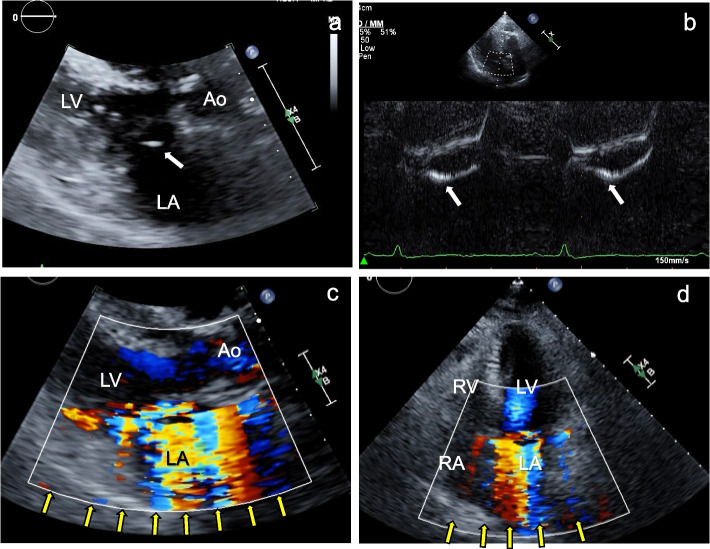
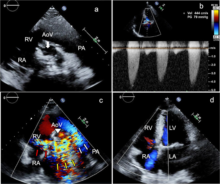
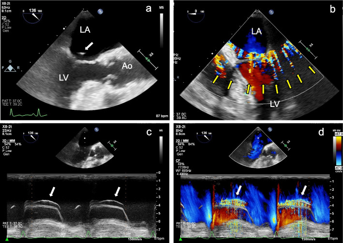
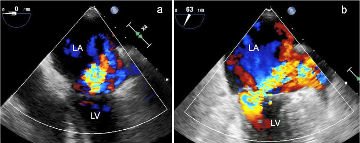
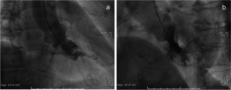
Case 1: An 85-year-old patient was referred for mitral valve surgery due to suspected severe mitral regurgitation (MR). Upon evaluation, transthoracic echocardiography (TTE) showed mitral valve prolapse (P3) and a high-echoic, vibrating structure attached to the mitral valve, indicative of chordal rupture. Color Doppler echocardiography revealed strong systolic signals in the left atrium, mimicking severe MR. Transesophageal echocardiography (TEE) also detected the vibrating structure and color Doppler stripes in the left atrium, left ventricle, and outside the cardiac chambers. The PISA method on TEE indicated moderate MR and left ventriculography showed Sellers grade II MR. The artifact was identified as color Doppler stripes caused by the vibrating high-echoic structure from the ruptured chorda. Case 2: A 64-year-old patient with severe aortic stenosis, end-stage kidney disease requiring hemodialysis, and a history of coronary bypass grafting presented for routine follow-up. B-mode echocardiography showed a severely calcified tricuspid aortic valve with a vibrating calcified nodule and restricted opening, corresponding to severe aortic stenosis. During systole, color Doppler signals were observed around the aortic, pulmonary, and tricuspid valves, mimicking significant pulmonary stenosis and tricuspid regurgitation. However, pulmonary stenosis was ruled out as the pulmonary valve opening was normal. Mild tricuspid regurgitation was confirmed in the apical view.

CONCLUSIONS

These cases highlight the diagnostic challenges posed by color Doppler stripes. Recognizing and understanding this artifact are crucial for the accurate diagnosis and management of VHD, ensuring appropriate treatment and patient outcomes.

摘要

背景

超声心动图仍然是评估瓣膜性心脏病(VHD)的参考标准成像技术,但像“彩色多普勒条纹”这样的伪影会使诊断变得复杂。这种伪影并未被广泛认识,可能会模拟严重的 VHD,导致潜在的误诊。我们报告了两个彩色多普勒条纹模拟严重 VHD 的病例,强调了在超声心动图评估中需要注意和准确解读。

病例介绍

病例 1:一位 85 岁的患者因疑似严重二尖瓣反流(MR)而被转诊接受二尖瓣手术。评估时,经胸超声心动图(TTE)显示二尖瓣脱垂(P3)和附着在二尖瓣上的高回声、振动结构,提示腱索断裂。彩色多普勒超声心动图显示左心房有强烈的收缩期信号,模拟严重的 MR。经食管超声心动图(TEE)也检测到左心房、左心室和心脏室外的振动结构和彩色多普勒条纹。TEE 上的 PISA 法提示中度 MR,左心室造影显示 Sellers 分级 II 级 MR。该伪影被识别为来自断裂腱索的振动高回声结构引起的彩色多普勒条纹。病例 2:一位 64 岁的患者,患有严重的主动脉瓣狭窄、终末期肾病需要血液透析,以及冠状动脉旁路移植术的病史,来进行常规随访。B 型超声心动图显示严重钙化的三尖瓣主动脉瓣,有一个振动的钙化结节和限制瓣口开放,对应严重的主动脉瓣狭窄。收缩期时,在主动脉瓣、肺动脉瓣和三尖瓣周围观察到彩色多普勒信号,模拟严重的肺动脉瓣狭窄和三尖瓣反流。然而,由于肺动脉瓣开口正常,排除了肺动脉瓣狭窄。在心尖观确认了轻度三尖瓣反流。

结论

这些病例强调了彩色多普勒条纹带来的诊断挑战。认识和理解这种伪影对于准确诊断和管理 VHD 至关重要,确保了适当的治疗和患者的预后。

https://cdn.ncbi.nlm.nih.gov/pmc/blobs/3b0f/11457407/67c62d80977d/12947_2024_331_Fig5_HTML.jpg
https://cdn.ncbi.nlm.nih.gov/pmc/blobs/3b0f/11457407/516dddc98a3e/12947_2024_331_Fig1_HTML.jpg
https://cdn.ncbi.nlm.nih.gov/pmc/blobs/3b0f/11457407/e8541ed9212e/12947_2024_331_Fig2_HTML.jpg
https://cdn.ncbi.nlm.nih.gov/pmc/blobs/3b0f/11457407/6c7060a90d34/12947_2024_331_Fig3_HTML.jpg
https://cdn.ncbi.nlm.nih.gov/pmc/blobs/3b0f/11457407/41e9eadd3225/12947_2024_331_Fig4_HTML.jpg
https://cdn.ncbi.nlm.nih.gov/pmc/blobs/3b0f/11457407/67c62d80977d/12947_2024_331_Fig5_HTML.jpg

相似文献

1
'Color Doppler stripes' make it difficult to diagnose the severity of valvular heart diseases: a report of two cases.“彩色多普勒条纹”使瓣膜性心脏病的严重程度难以诊断:两例报告。
Cardiovasc Ultrasound. 2024 Oct 7;22(1):12. doi: 10.1186/s12947-024-00331-1.
2
Identifying the Severity of Heart Valve Stenosis and Regurgitation Among a Diverse Population Within an Integrated Health Care System: Natural Language Processing Approach.在综合性医疗保健系统中识别不同人群中心脏瓣膜狭窄和反流的严重程度:自然语言处理方法。
JMIR Cardio. 2024 Sep 30;8:e60503. doi: 10.2196/60503.
3
Echocardiographic assessment of mitral stenosis and its associated valvular lesions in 205 patients and lack of association with mitral valve prolapse.205例二尖瓣狭窄及其相关瓣膜病变的超声心动图评估以及与二尖瓣脱垂的无关性
J Am Soc Echocardiogr. 1997 Mar;10(2):141-8. doi: 10.1016/s0894-7317(97)70086-5.
4
[Value of transesophageal echocardiography in diagnosis of diseases of native heart valves].经食管超声心动图在诊断天然心脏瓣膜疾病中的价值
Herz. 1993 Dec;18(6):329-40.
5
Clinical and echocardiographic evaluation of mitral valve aneurysms: a retrospective, single center study.临床和超声心动图评估二尖瓣动脉瘤:一项回顾性、单中心研究。
Int J Cardiovasc Imaging. 2014 Mar;30(3):535-41. doi: 10.1007/s10554-014-0365-4. Epub 2014 Jan 14.
6
Transesophageal echocardiographic (TEE) evaluation of the mitral and tricuspid valves.经食管超声心动图(TEE)对二尖瓣和三尖瓣的评估。
Cardiol Clin. 2000 Nov;18(4):731-50. doi: 10.1016/s0733-8651(05)70177-6.
7
Biplane transesophageal color Doppler echocardiography for assessment of mitral valve area with mitral inflow jet widths.双平面经食管彩色多普勒超声心动图通过二尖瓣血流喷射宽度评估二尖瓣面积。
J Am Soc Echocardiogr. 1995 Mar-Apr;8(2):121-31. doi: 10.1016/s0894-7317(05)80401-8.
8
Three-dimensional color Doppler reconstruction of intracardiac blood flow in patients with different heart valve diseases.不同心脏瓣膜疾病患者心腔内血流的三维彩色多普勒重建
Am J Cardiol. 2000 Dec 15;86(12):1343-8. doi: 10.1016/s0002-9149(00)01239-x.
9
Role of echocardiography for catheter-based management of valvular heart disease.超声心动图在基于导管的心脏瓣膜病治疗中的作用。
J Cardiol. 2017 Jan;69(1):66-73. doi: 10.1016/j.jjcc.2016.09.015. Epub 2016 Nov 15.
10
A prospective echocardiographic study of the effects of balloon mitral commissurotomy on pre-existing mitral regurgitation in patients with mitral stenosis.一项关于球囊二尖瓣交界切开术对二尖瓣狭窄患者原有二尖瓣反流影响的前瞻性超声心动图研究。
Cardiology. 1998 Mar;89(3):202-9. doi: 10.1159/000006788.

本文引用的文献

1
Color Doppler Splay in Mitral Regurgitation: Hemodynamic Correlates and Outcomes in a Clinical Cohort.二尖瓣反流中的彩色多普勒分流:临床队列中的血流动力学关联及结局
J Am Soc Echocardiogr. 2022 Sep;35(9):933-939. doi: 10.1016/j.echo.2022.04.006. Epub 2022 Apr 23.
2
2021 ESC/EACTS Guidelines for the management of valvular heart disease.2021年欧洲心脏病学会/欧洲心胸外科学会瓣膜性心脏病管理指南。
Eur Heart J. 2022 Feb 12;43(7):561-632. doi: 10.1093/eurheartj/ehab395.
3
Global epidemiology of valvular heart disease.全球瓣膜性心脏病的流行病学。
Nat Rev Cardiol. 2021 Dec;18(12):853-864. doi: 10.1038/s41569-021-00570-z. Epub 2021 Jun 25.
4
Response to: "Color Doppler Splay: A New Tool for the Assessment of Valvular Regurgitations?" by Allievi et al.对阿列维等人所著《彩色多普勒散开:评估瓣膜反流的新工具?》的回应
J Am Soc Echocardiogr. 2021 Sep;34(9):1022-1023. doi: 10.1016/j.echo.2021.05.014. Epub 2021 May 29.
5
Color Doppler Splay: A New Tool for the Assessment of Valvular Regurgitations?彩色多普勒展开:评估瓣膜反流的新工具?
J Am Soc Echocardiogr. 2021 Sep;34(9):1021-1022. doi: 10.1016/j.echo.2021.05.003. Epub 2021 May 11.
6
2020 ACC/AHA Guideline for the Management of Patients With Valvular Heart Disease: A Report of the American College of Cardiology/American Heart Association Joint Committee on Clinical Practice Guidelines.2020美国心脏病学会/美国心脏协会瓣膜性心脏病患者管理指南:美国心脏病学会/美国心脏协会临床实践指南联合委员会报告
Circulation. 2021 Feb 2;143(5):e72-e227. doi: 10.1161/CIR.0000000000000923. Epub 2020 Dec 17.
7
JCS/JSCS/JATS/JSVS 2020 Guidelines on the Management of Valvular Heart Disease.JCS/JSCS/JATS/JSVS 2020年心脏瓣膜病管理指南
Circ J. 2020 Oct 23;84(11):2037-2119. doi: 10.1253/circj.CJ-20-0135. Epub 2020 Sep 11.
8
Color Doppler Splay: A Clue to the Presence of Significant Mitral Regurgitation.彩色多谱勒分流:提示存在显著二尖瓣反流。
J Am Soc Echocardiogr. 2020 Oct;33(10):1212-1219.e1. doi: 10.1016/j.echo.2020.05.002. Epub 2020 Jul 22.
9
Echocardiography in Transcatheter Structural Heart Disease Interventions.经导管结构性心脏病介入治疗中的超声心动图。
Prog Cardiovasc Dis. 2018 Nov-Dec;61(5-6):423-436. doi: 10.1016/j.pcad.2018.11.009. Epub 2018 Nov 20.
10
Ultrasound imaging artifacts: How to recognize them and how to avoid them.超声成像伪像:如何识别它们以及如何避免它们。
Echocardiography. 2018 Sep;35(9):1388-1401. doi: 10.1111/echo.14116. Epub 2018 Aug 6.