Adesoye Samuel, Al Abdullah Saqer, Kumari Anjali, Pathiraja Gayani, Nowlin Kyle, Dellinger Kristen
Department of Nanoengineering, Joint School of Nanoscience and Nanoengineering, North Carolina A&T State University, 2907 E Gate City Blvd, Greensboro, NC 27401, USA.
Department of Nanoscience, Joint School of Nanoscience and Nanoengineering, University of North Carolina at Greensboro, 2907 E Gate City Blvd, Greensboro, NC 27401, USA.
Chemosensors (Basel). 2023 Nov;11(11). doi: 10.3390/chemosensors11110554. Epub 2023 Nov 5.
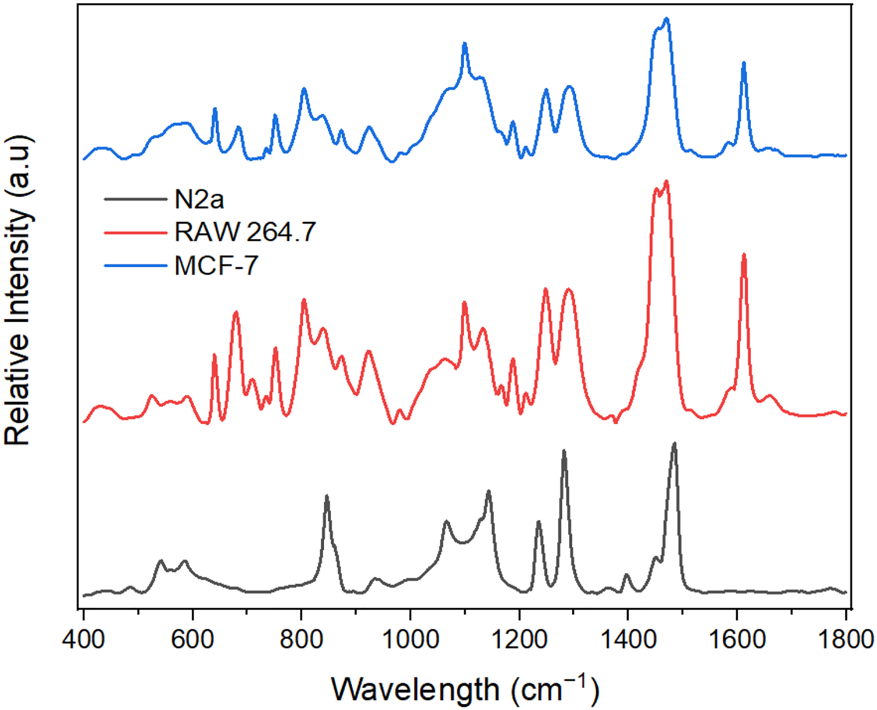
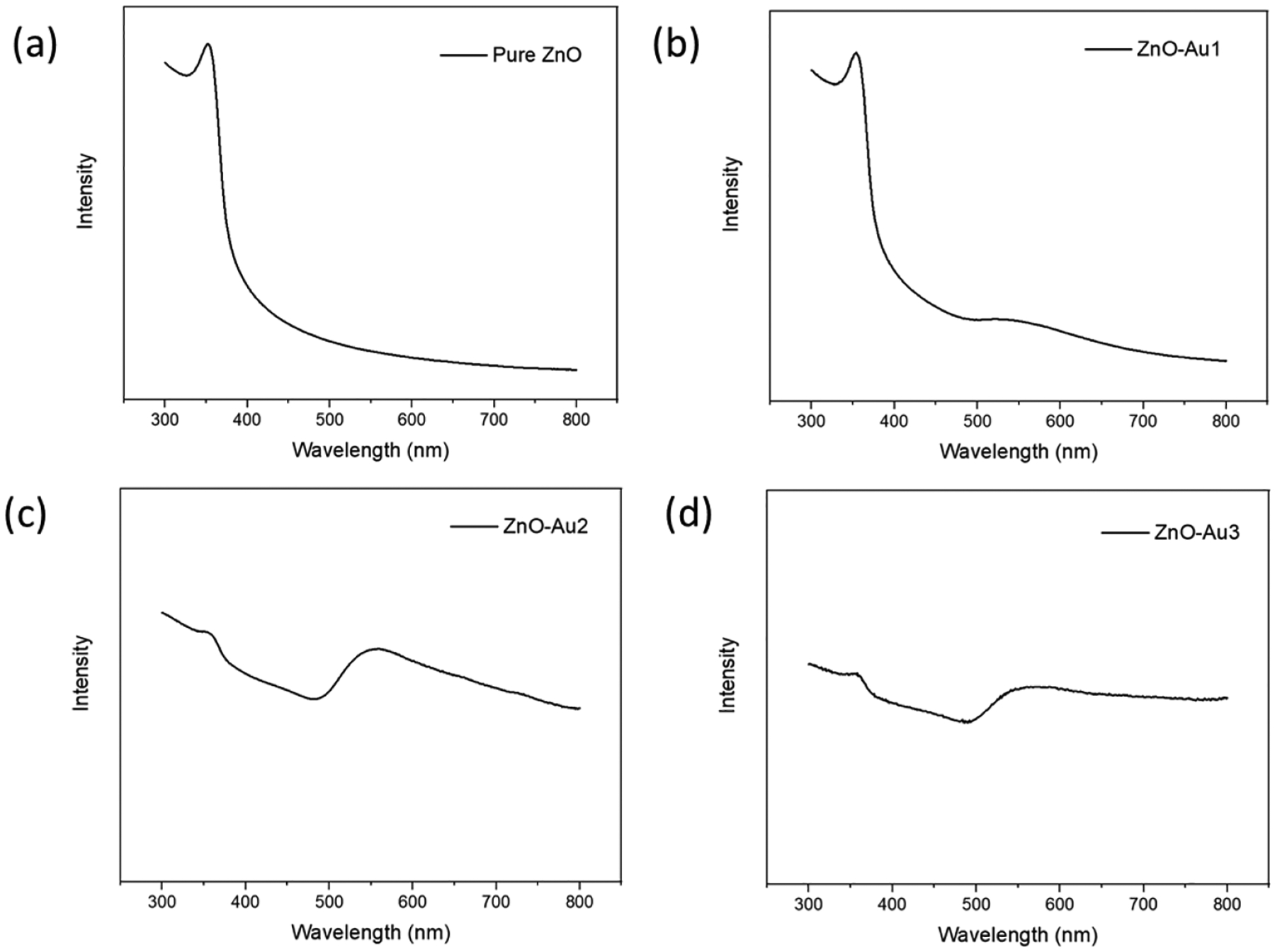
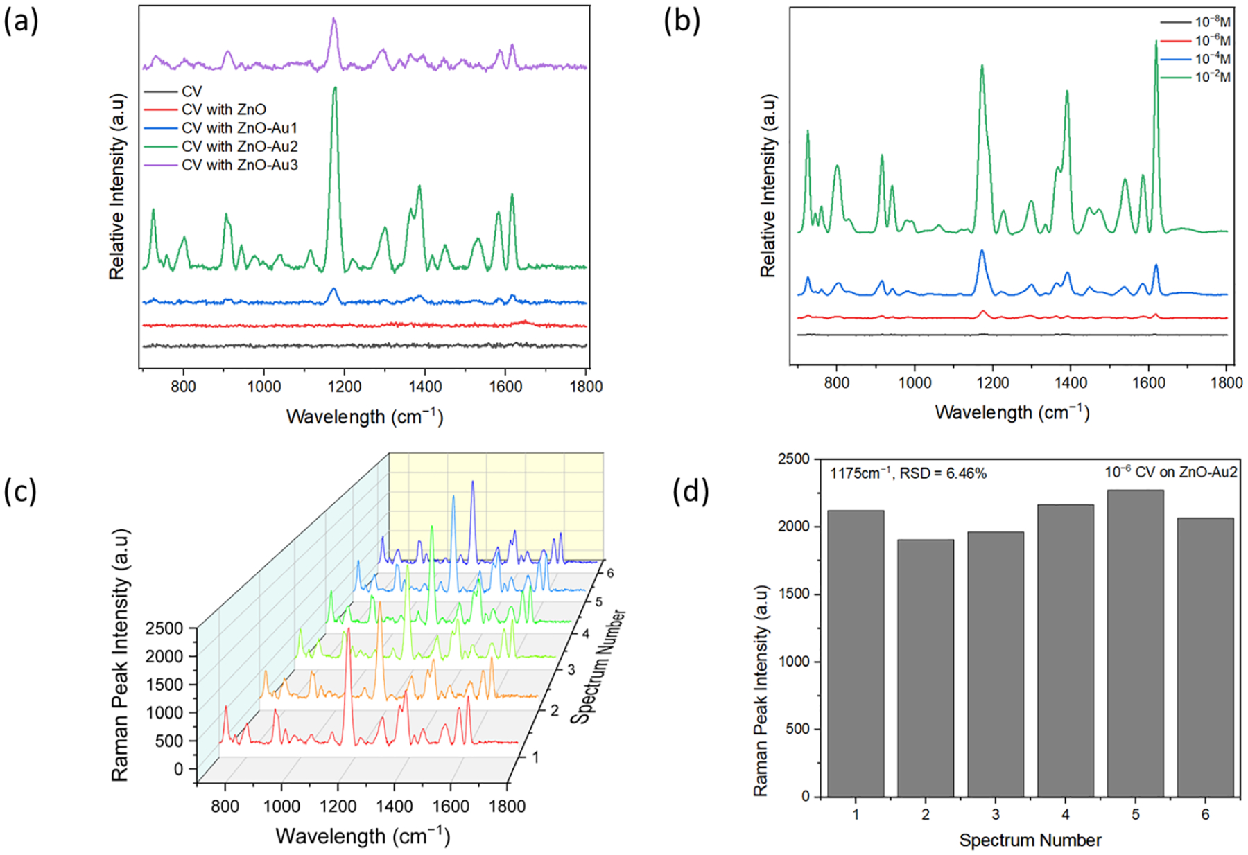
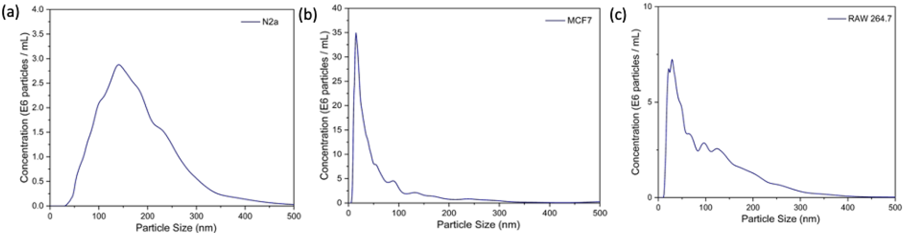
Developing a biomolecular detection method that minimizes photodamage while preserving an environment suitable for biological constituents to maintain their physiological state is expected to drive new diagnostic and mechanistic breakthroughs. In addition, ultra-sensitive diagnostic platforms are needed for rapid and point-of-care technologies for various diseases. Considering this, surface-enhanced Raman scattering (SERS) is proposed as a non-destructive and sensitive approach to address the limitations of fluorescence, electrochemical, and other optical detection techniques. However, to advance the applications of SERS, novel approaches that can enhance the signal of substrate materials are needed to improve reproducibility and costs associated with manufacture and scale-up. Due to their physical properties and synthesis, semiconductor-based nanostructures have gained increasing recognition as SERS substrates; however, low signal enhancements have offset their widespread adoption. To address this limitation and assess the potential for use in biological applications, zinc oxide (ZnO) was coated with different concentrations (0.01-0.1 M) of gold (Au) precursor. When crystal violet (CV) was used as a model target with the synthesized substrates, the highest enhancement was obtained with ZnO coated with 0.05 M Au precursor. This substrate was subsequently applied to differentiate exosomes derived from three cell types to provide insight into their molecular diversity. We anticipate this work will serve as a platform for colloidal hybrid SERS substrates in future bio-sensing applications.
开发一种生物分子检测方法,在保留适合生物成分维持其生理状态的环境的同时尽量减少光损伤,有望推动新的诊断和机制突破。此外,各种疾病的快速即时检测技术需要超灵敏的诊断平台。考虑到这一点,表面增强拉曼散射(SERS)被提议作为一种无损且灵敏的方法,以解决荧光、电化学和其他光学检测技术的局限性。然而,为了推进SERS的应用,需要能够增强基底材料信号的新方法,以提高与制造和扩大规模相关的重现性和成本。由于其物理性质和合成方法,基于半导体的纳米结构作为SERS基底越来越受到认可;然而,低信号增强抵消了它们的广泛应用。为了解决这一局限性并评估其在生物应用中的潜力,用不同浓度(0.01 - 0.1 M)的金(Au)前驱体包覆氧化锌(ZnO)。当使用结晶紫(CV)作为合成基底的模型目标时,用0.05 M Au前驱体包覆的ZnO获得了最高的增强效果。随后将该基底应用于区分源自三种细胞类型的外泌体,以深入了解它们的分子多样性。我们预计这项工作将成为未来生物传感应用中胶体混合SERS基底的一个平台。