• 文献检索
  • 文档翻译
  • 深度研究
  • 学术资讯
  • Suppr Zotero 插件Zotero 插件
  • 邀请有礼
  • 套餐&价格
  • 历史记录
应用&插件
Suppr Zotero 插件Zotero 插件浏览器插件Mac 客户端Windows 客户端微信小程序
定价
高级版会员购买积分包购买API积分包
服务
文献检索文档翻译深度研究API 文档MCP 服务
关于我们
关于 Suppr公司介绍联系我们用户协议隐私条款
关注我们

Suppr 超能文献

核心技术专利:CN118964589B侵权必究
粤ICP备2023148730 号-1Suppr @ 2026

文献检索

告别复杂PubMed语法,用中文像聊天一样搜索,搜遍4000万医学文献。AI智能推荐,让科研检索更轻松。

立即免费搜索

文件翻译

保留排版,准确专业,支持PDF/Word/PPT等文件格式,支持 12+语言互译。

免费翻译文档

深度研究

AI帮你快速写综述,25分钟生成高质量综述,智能提取关键信息,辅助科研写作。

立即免费体验

胚胎植入前小鼠的核实例分割和跟踪。

Nuclear instance segmentation and tracking for preimplantation mouse embryos.

机构信息

Center for Computational Biology, Flatiron Institute - Simons Foundation, New York, NY 10010, USA.

Department of Chemical and Biological Engineering, Princeton University, Princeton, NJ 08544, USA.

出版信息

Development. 2024 Nov 1;151(21). doi: 10.1242/dev.202817. Epub 2024 Nov 6.

DOI:10.1242/dev.202817
PMID:39373366
原文链接:https://pmc.ncbi.nlm.nih.gov/articles/PMC11574361/
Abstract

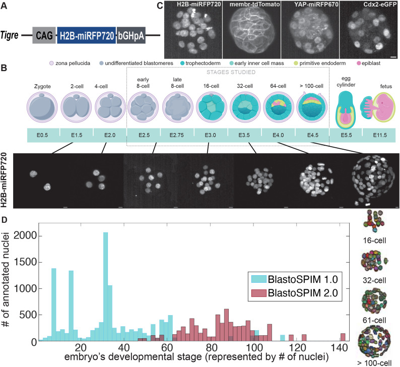
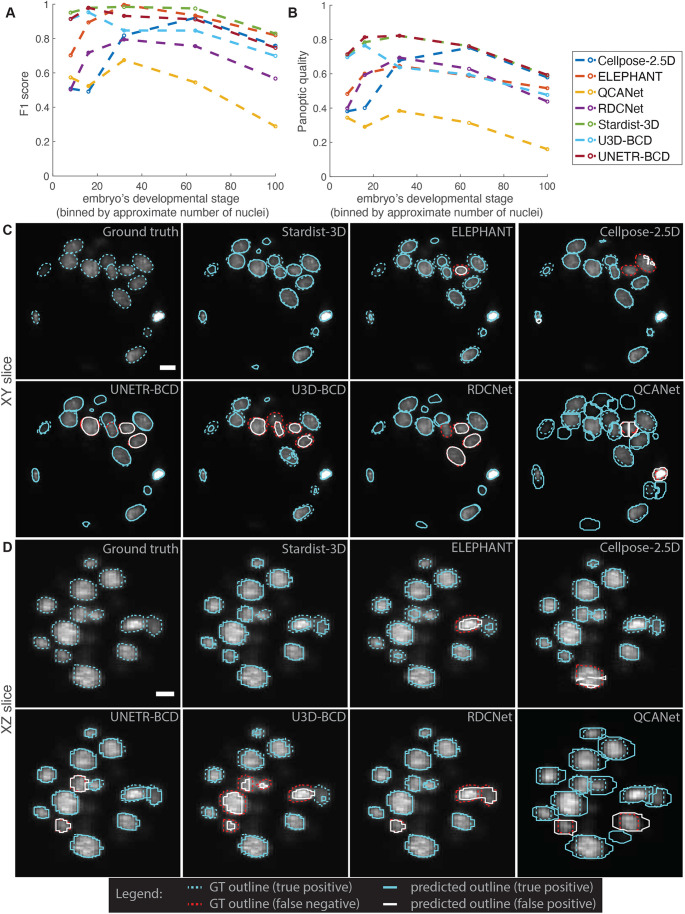
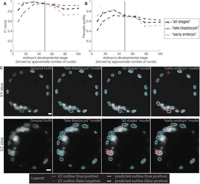
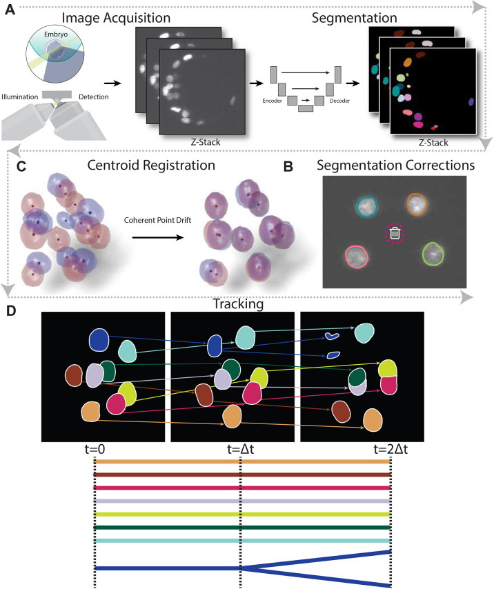
For investigations into fate specification and morphogenesis in time-lapse images of preimplantation embryos, automated 3D instance segmentation and tracking of nuclei are invaluable. Low signal-to-noise ratio, high voxel anisotropy, high nuclear density, and variable nuclear shapes can limit the performance of segmentation methods, while tracking is complicated by cell divisions, low frame rates, and sample movements. Supervised machine learning approaches can radically improve segmentation accuracy and enable easier tracking, but they often require large amounts of annotated 3D data. Here, we first report a previously unreported mouse line expressing near-infrared nuclear reporter H2B-miRFP720. We then generate a dataset (termed BlastoSPIM) of 3D images of H2B-miRFP720-expressing embryos with ground truth for nuclear instances. Using BlastoSPIM, we benchmark seven convolutional neural networks and identify Stardist-3D as the most accurate instance segmentation method. With our BlastoSPIM-trained Stardist-3D models, we construct a complete pipeline for nuclear instance segmentation and lineage tracking from the eight-cell stage to the end of preimplantation development (>100 nuclei). Finally, we demonstrate the usefulness of BlastoSPIM as pre-train data for related problems, both for a different imaging modality and for different model systems.

摘要

对于在胚胎植入前的延时图像中进行命运特化和形态发生的研究,自动化的 3D 实例分割和核追踪是非常宝贵的。低信噪比、高体素各向异性、高核密度和可变的核形状会限制分割方法的性能,而追踪则受到细胞分裂、低帧率和样本运动的影响。监督机器学习方法可以极大地提高分割准确性并实现更简单的追踪,但它们通常需要大量的标注 3D 数据。在这里,我们首先报告了一个以前未报道的表达近红外核报告基因 H2B-miRFP720 的小鼠品系。然后,我们生成了一个具有核实例真实数据的 H2B-miRFP720 表达胚胎的 3D 图像数据集(称为 BlastoSPIM)。使用 BlastoSPIM,我们对七个卷积神经网络进行了基准测试,并确定 Stardist-3D 是最准确的实例分割方法。使用我们的 BlastoSPIM 训练的 Stardist-3D 模型,我们构建了一个从八细胞阶段到植入前发育结束(>100 个核)的核实例分割和谱系追踪的完整流水线。最后,我们展示了 BlastoSPIM 作为相关问题的预训练数据的有用性,无论是对于不同的成像模式还是对于不同的模型系统。

https://cdn.ncbi.nlm.nih.gov/pmc/blobs/3ade/11574361/eba4f4ce97b1/develop-151-202817-g6.jpg
https://cdn.ncbi.nlm.nih.gov/pmc/blobs/3ade/11574361/166f84a98e79/develop-151-202817-g1.jpg
https://cdn.ncbi.nlm.nih.gov/pmc/blobs/3ade/11574361/c4c6489d28c4/develop-151-202817-g2.jpg
https://cdn.ncbi.nlm.nih.gov/pmc/blobs/3ade/11574361/952a235e2b44/develop-151-202817-g3.jpg
https://cdn.ncbi.nlm.nih.gov/pmc/blobs/3ade/11574361/8c03548e7e0b/develop-151-202817-g4.jpg
https://cdn.ncbi.nlm.nih.gov/pmc/blobs/3ade/11574361/c868b46c6ca8/develop-151-202817-g5.jpg
https://cdn.ncbi.nlm.nih.gov/pmc/blobs/3ade/11574361/eba4f4ce97b1/develop-151-202817-g6.jpg

相似文献

1
Nuclear instance segmentation and tracking for preimplantation mouse embryos.胚胎植入前小鼠的核实例分割和跟踪。
Development. 2024 Nov 1;151(21). doi: 10.1242/dev.202817. Epub 2024 Nov 6.
2
A novel ground truth dataset enables robust 3D nuclear instance segmentation in early mouse embryos.一个全新的地面真值数据集助力早期小鼠胚胎中稳健的三维细胞核实例分割。
bioRxiv. 2023 Mar 15:2023.03.14.532646. doi: 10.1101/2023.03.14.532646.
3
Automatic three-dimensional segmentation of mouse embryonic stem cell nuclei by utilising multiple channels of confocal fluorescence images.利用共聚焦荧光图像的多个通道自动对小鼠胚胎干细胞核进行三维分割。
J Microsc. 2021 Jan;281(1):57-75. doi: 10.1111/jmi.12949. Epub 2020 Aug 8.
4
A deep learning-based toolkit for 3D nuclei segmentation and quantitative analysis in cellular and tissue context.基于深度学习的 3D 细胞核分割和细胞及组织定量分析工具包。
Development. 2024 Jul 15;151(14). doi: 10.1242/dev.202800. Epub 2024 Jul 18.
5
3D convolutional neural networks-based segmentation to acquire quantitative criteria of the nucleus during mouse embryogenesis.基于 3D 卷积神经网络的分割技术,获取小鼠胚胎发生过程中细胞核的定量标准。
NPJ Syst Biol Appl. 2020 Oct 20;6(1):32. doi: 10.1038/s41540-020-00152-8.
6
Bi-channel image registration and deep-learning segmentation (BIRDS) for efficient, versatile 3D mapping of mouse brain.双通道图像配准和深度学习分割(BIRDS)用于高效、通用的小鼠大脑 3D 映射。
Elife. 2021 Jan 18;10:e63455. doi: 10.7554/eLife.63455.
7
Cell cycle phase classification in 3D in vivo microscopy of Drosophila embryogenesis.果蝇胚胎发生的三维活体显微镜中的细胞周期相分类。
BMC Bioinformatics. 2011;12 Suppl 13(Suppl 13):S18. doi: 10.1186/1471-2105-12-S13-S18. Epub 2011 Nov 30.
8
A novel cell nuclei segmentation method for 3D C. elegans embryonic time-lapse images.一种用于 3D C. elegans 胚胎延时图像的新型细胞核分割方法。
BMC Bioinformatics. 2013 Nov 19;14:328. doi: 10.1186/1471-2105-14-328.
9
Single-cell segmentation in bacterial biofilms with an optimized deep learning method enables tracking of cell lineages and measurements of growth rates.利用优化的深度学习方法对细菌生物膜中的单细胞进行分割,可实现细胞谱系追踪和生长速率测量。
Mol Microbiol. 2023 Jun;119(6):659-676. doi: 10.1111/mmi.15064. Epub 2023 Apr 17.
10
Semi-supervised learning for automatic segmentation of the knee from MRI with convolutional neural networks.基于卷积神经网络的膝关节 MRI 半自动分割的半监督学习。
Comput Methods Programs Biomed. 2020 Jun;189:105328. doi: 10.1016/j.cmpb.2020.105328. Epub 2020 Jan 11.

引用本文的文献

1
Generative model for the first cell fate bifurcation in mammalian development.哺乳动物发育中首个细胞命运分支的生成模型。
Development. 2025 Aug 5. doi: 10.1242/dev.204717.
2
Live imaging endogenous transcription factor dynamics reveals mechanisms of epiblast and primitive endoderm fate segregation.实时成像内源性转录因子动力学揭示上胚层和原始内胚层命运分离的机制。
Curr Biol. 2025 Jul 26. doi: 10.1016/j.cub.2025.07.031.
3
Geometric, cell cycle and maternal-to-zygotic transition-associated YAP dynamics during preimplantation embryo development.

本文引用的文献

1
A deep learning-based toolkit for 3D nuclei segmentation and quantitative analysis in cellular and tissue context.基于深度学习的 3D 细胞核分割和细胞及组织定量分析工具包。
Development. 2024 Jul 15;151(14). doi: 10.1242/dev.202800. Epub 2024 Jul 18.
2
Segmentation metric misinterpretations in bioimage analysis.生物影像分析中的分割度量误读。
Nat Methods. 2024 Feb;21(2):213-216. doi: 10.1038/s41592-023-01942-8. Epub 2023 Jul 27.
3
Nuclear compression regulates YAP spatiotemporal fluctuations in living cells.核压缩调节活细胞中 YAP 的时空波动。
着床前胚胎发育过程中与几何形状、细胞周期和母源-合子转变相关的YAP动态变化。
Dev Biol. 2025 Aug;524:105-115. doi: 10.1016/j.ydbio.2025.05.006. Epub 2025 May 9.
4
A model for boundary-driven tissue morphogenesis.一种边界驱动组织形态发生的模型。
ArXiv. 2025 Mar 5:arXiv:2503.03688v1.
5
Generative model for the first cell fate bifurcation in mammalian development.哺乳动物发育中首个细胞命运分支的生成模型。
bioRxiv. 2025 Feb 25:2025.02.24.639895. doi: 10.1101/2025.02.24.639895.
6
Geometric, cell cycle and maternal-to-zygotic transition-associated YAP dynamics during preimplantation embryo development.着床前胚胎发育过程中与几何形状、细胞周期和母源-合子转变相关的YAP动态变化
bioRxiv. 2025 Feb 27:2025.02.27.640568. doi: 10.1101/2025.02.27.640568.
7
A tug-of-war between germ cell motility and intercellular bridges controls germline cyst formation in mice.生殖细胞运动性与细胞间桥之间的拔河比赛控制着小鼠生殖系囊肿的形成。
Curr Biol. 2024 Dec 16;34(24):5728-5738.e4. doi: 10.1016/j.cub.2024.10.062. Epub 2024 Nov 19.
Proc Natl Acad Sci U S A. 2023 Jul 11;120(28):e2301285120. doi: 10.1073/pnas.2301285120. Epub 2023 Jul 3.
4
Multiscale light-sheet organoid imaging framework.多尺度光片器官成像框架。
Nat Commun. 2022 Aug 18;13(1):4864. doi: 10.1038/s41467-022-32465-z.
5
The Nuclear-to-Cytoplasmic Ratio: Coupling DNA Content to Cell Size, Cell Cycle, and Biosynthetic Capacity.核质比:将 DNA 含量与细胞大小、细胞周期和生物合成能力相偶联。
Annu Rev Genet. 2022 Nov 30;56:165-185. doi: 10.1146/annurev-genet-080320-030537. Epub 2022 Aug 17.
6
EmbedSeg: Embedding-based Instance Segmentation for Biomedical Microscopy Data.EmbedSeg:基于嵌入的生物医学显微镜数据实例分割。
Med Image Anal. 2022 Oct;81:102523. doi: 10.1016/j.media.2022.102523. Epub 2022 Jul 3.
7
GIANI - open-source software for automated analysis of 3D microscopy images.GIANI - 用于 3D 显微镜图像自动分析的开源软件。
J Cell Sci. 2022 May 15;135(10). doi: 10.1242/jcs.259511. Epub 2022 May 17.
8
Cell size and polarization determine cytokinesis furrow ingression dynamics in mouse embryos.细胞大小和极化决定了小鼠胚胎胞质分裂沟内陷的动力学。
Proc Natl Acad Sci U S A. 2022 Mar 22;119(12):e2119381119. doi: 10.1073/pnas.2119381119. Epub 2022 Mar 16.
9
Live imaging YAP signalling in mouse embryo development.活细胞成像观察 YAP 信号在小鼠胚胎发育中的作用。
Open Biol. 2022 Jan;12(1):210335. doi: 10.1098/rsob.210335. Epub 2022 Jan 19.
10
Tracking cell lineages in 3D by incremental deep learning.通过渐进式深度学习追踪 3D 中的细胞谱系。
Elife. 2022 Jan 6;11:e69380. doi: 10.7554/eLife.69380.