Zhao Xiao, Dong Yue-Han, Xu Li-Yu, Shen Yan-Yan, Qin Gang, Zhang Zheng-Bo
Department of Applied Engineering, Zhejiang Institute of Economics and Trade, Hangzhou, Zhejiang Province, 310018, China.
Wuxi Hospital of Traditional Chinese Medicine, Wuxi, Jiangsu Province, 214071, China.
J Bone Oncol. 2024 Sep 25;48:100638. doi: 10.1016/j.jbo.2024.100638. eCollection 2024 Oct.
The objective of this study is to develop a novel diagnostic tool using deep learning and radiomics to distinguish bone tumors on CT images as metastases from breast cancer. By providing a more accurate and reliable method for identifying metastatic bone tumors, this approach aims to significantly improve clinical decision-making and patient management in the context of breast cancer.
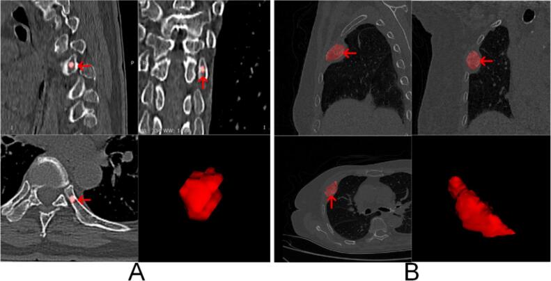
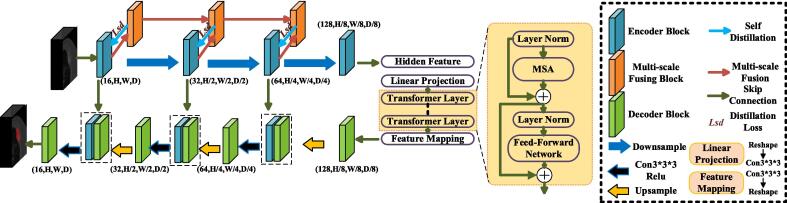
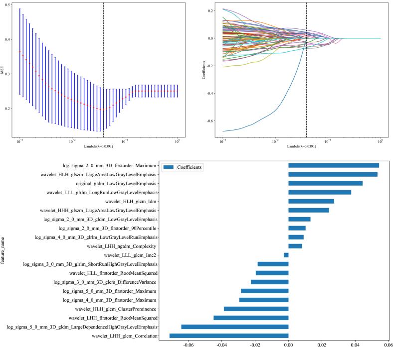
This study utilized CT images of bone tumors from 178 patients, including 78 cases of breast cancer bone metastases and 100 cases of non-breast cancer bone metastases. The dataset was processed using the Medical Image Segmentation via Self-distilling TransUNet (MISSU) model for automated segmentation. Radiomics features were extracted from the segmented tumor regions using the Pyradiomics library, capturing various aspects of tumor phenotype. Feature selection was conducted using LASSO regression to identify the most predictive features. The model's performance was evaluated using ten-fold cross-validation, with metrics including accuracy, sensitivity, specificity, and the Dice similarity coefficient.
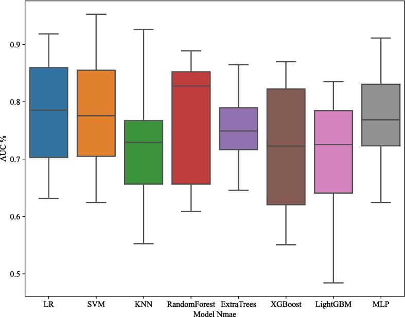
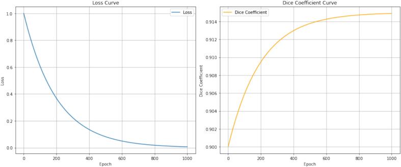
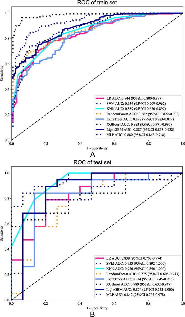
The developed radiomics model using the SVM algorithm achieved high discriminatory power, with an AUC of 0.936 on the training set and 0.953 on the test set. The model's performance metrics demonstrated strong accuracy, sensitivity, and specificity. Specifically, the accuracy was 0.864 for the training set and 0.853 for the test set. Sensitivity values were 0.838 and 0.789 for the training and test sets, respectively, while specificity values were 0.896 and 0.933 for the training and test sets, respectively. These results indicate that the SVM model effectively distinguishes between bone metastases originating from breast cancer and other origins. Additionally, the average Dice similarity coefficient for the automated segmentation was 0.915, demonstrating a high level of agreement with manual segmentations.
This study demonstrates the potential of combining CT-based radiomics and deep learning for the accurate detection of bone metastases from breast cancer. The high-performance metrics indicate that this approach can significantly enhance diagnostic accuracy, aiding in early detection and improving patient outcomes. Future research should focus on validating these findings on larger datasets, integrating the model into clinical workflows, and exploring its use in personalized treatment planning.
本研究的目的是开发一种利用深度学习和放射组学的新型诊断工具,以在CT图像上区分骨肿瘤是否为乳腺癌转移灶。通过提供一种更准确、可靠的方法来识别转移性骨肿瘤,该方法旨在显著改善乳腺癌背景下的临床决策和患者管理。
本研究使用了178例患者的骨肿瘤CT图像,其中包括78例乳腺癌骨转移患者和100例非乳腺癌骨转移患者。数据集通过基于自蒸馏TransUNet的医学图像分割(MISSU)模型进行处理,以实现自动分割。使用Pyradiomics库从分割后的肿瘤区域提取放射组学特征,捕捉肿瘤表型的各个方面。使用LASSO回归进行特征选择,以识别最具预测性的特征。使用十折交叉验证评估模型的性能,指标包括准确率、灵敏度、特异性和骰子相似系数。
使用支持向量机(SVM)算法开发的放射组学模型具有较高的鉴别力,训练集的AUC为0.936,测试集的AUC为0.953。模型的性能指标显示出较高的准确率、灵敏度和特异性。具体而言,训练集的准确率为0.864,测试集的准确率为0.853。训练集和测试集的灵敏度值分别为0.838和0.789,而训练集和测试集的特异性值分别为0.896和0.933。这些结果表明,SVM模型能够有效区分源自乳腺癌和其他来源的骨转移灶。此外,自动分割的平均骰子相似系数为0.915,表明与手动分割具有高度一致性。
本研究证明了将基于CT的放射组学与深度学习相结合用于准确检测乳腺癌骨转移灶的潜力。高性能指标表明,这种方法可以显著提高诊断准确性,有助于早期检测并改善患者预后。未来的研究应集中在更大的数据集上验证这些发现,将模型整合到临床工作流程中,并探索其在个性化治疗计划中的应用。