• 文献检索
  • 文档翻译
  • 深度研究
  • 学术资讯
  • Suppr Zotero 插件Zotero 插件
  • 邀请有礼
  • 套餐&价格
  • 历史记录
应用&插件
Suppr Zotero 插件Zotero 插件浏览器插件Mac 客户端Windows 客户端微信小程序
定价
高级版会员购买积分包购买API积分包
服务
文献检索文档翻译深度研究API 文档MCP 服务
关于我们
关于 Suppr公司介绍联系我们用户协议隐私条款
关注我们

Suppr 超能文献

核心技术专利:CN118964589B侵权必究
粤ICP备2023148730 号-1Suppr @ 2026

文献检索

告别复杂PubMed语法,用中文像聊天一样搜索,搜遍4000万医学文献。AI智能推荐,让科研检索更轻松。

立即免费搜索

文件翻译

保留排版,准确专业,支持PDF/Word/PPT等文件格式,支持 12+语言互译。

免费翻译文档

深度研究

AI帮你快速写综述,25分钟生成高质量综述,智能提取关键信息,辅助科研写作。

立即免费体验

一种从计算机断层扫描(CT)图像中进行半自动膝关节骨分割与重建的方法框架。

A method framework of semi-automatic knee bone segmentation and reconstruction from computed tomography (CT) images.

作者信息

Humayun Ahsan, Rehman Mustafain, Liu Bin

机构信息

International School of Information Science & Engineering (DUT-RUISE), Dalian University of Technology, Dalian, China.

Key Lab of Ubiquitous Network and Service Software of Liaoning Province, Dalian University of Technology, Dalian, China.

出版信息

Quant Imaging Med Surg. 2024 Oct 1;14(10):7151-7175. doi: 10.21037/qims-24-821. Epub 2024 Sep 26.

DOI:10.21037/qims-24-821
PMID:39429595
原文链接:https://pmc.ncbi.nlm.nih.gov/articles/PMC11485355/
Abstract

BACKGROUND

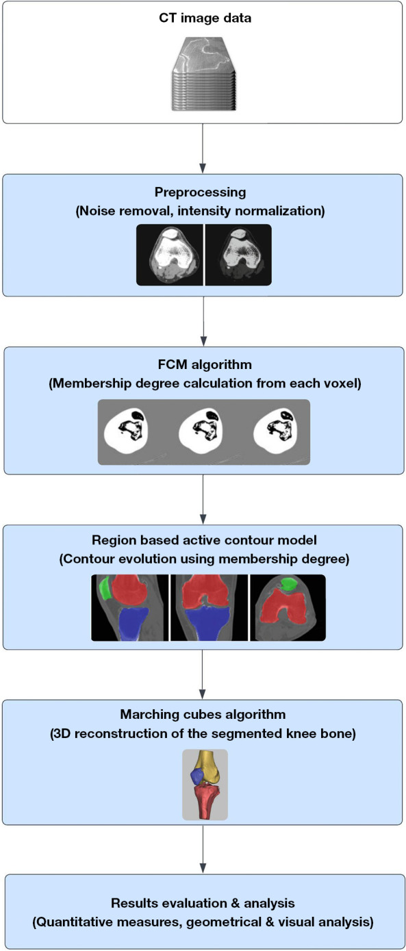
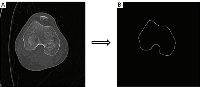
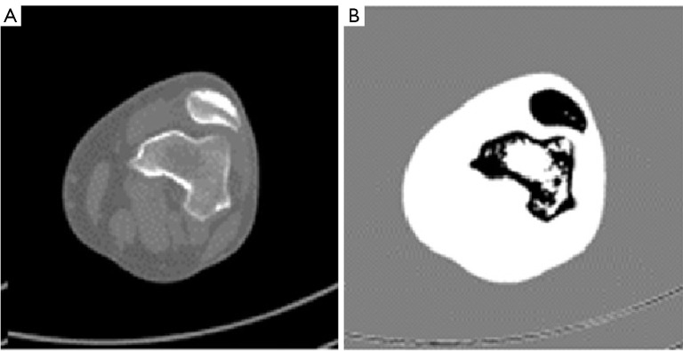
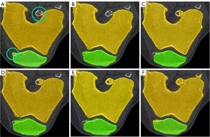
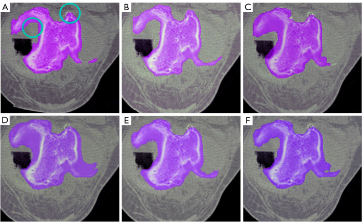
Accurate delineation of knee bone boundaries is crucial for computer-aided diagnosis (CAD) and effective treatment planning in knee diseases. Current methods often struggle with precise segmentation due to the knee joint's complexity, which includes intricate bone structures and overlapping soft tissues. These challenges are further complicated by variations in patient anatomy and image quality, highlighting the need for improved techniques. This paper presents a novel semi-automatic segmentation method for extracting knee bones from sequential computed tomography (CT) images.

METHODS

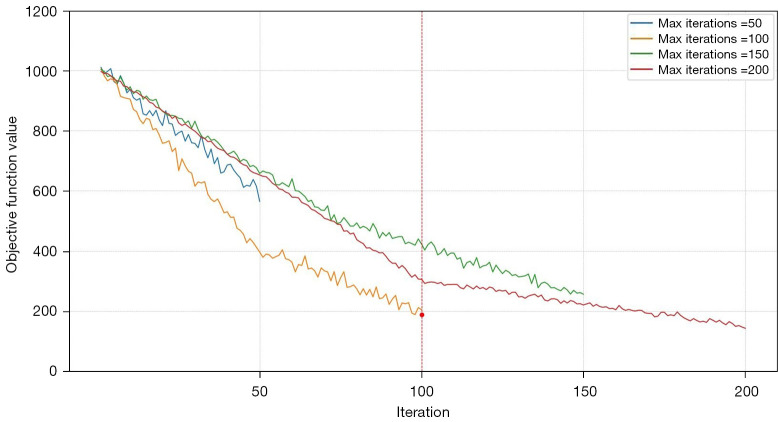
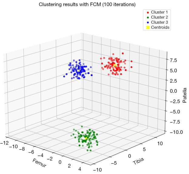
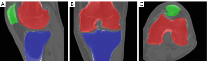
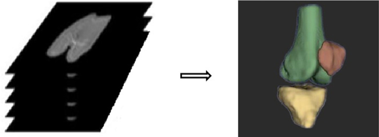
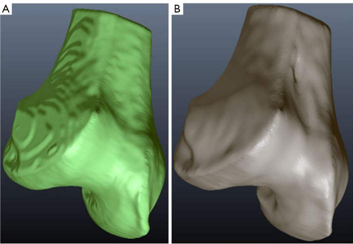
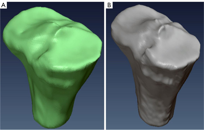
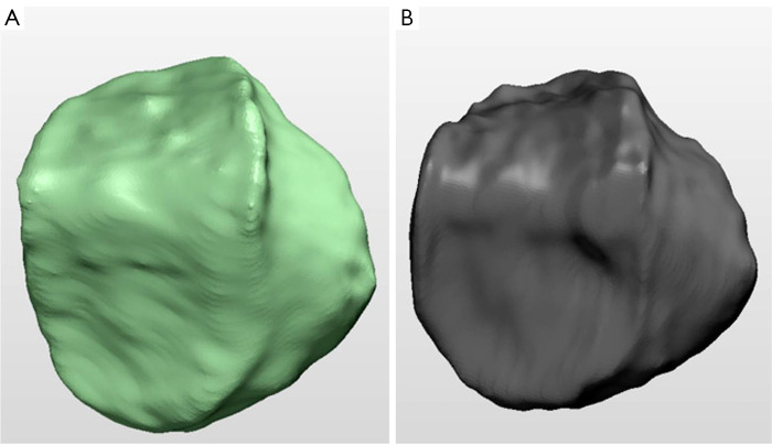
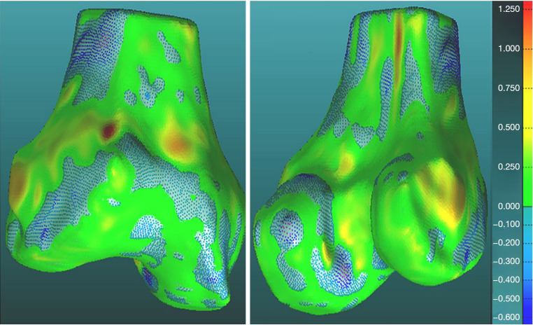
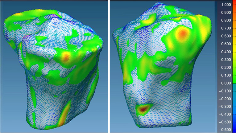
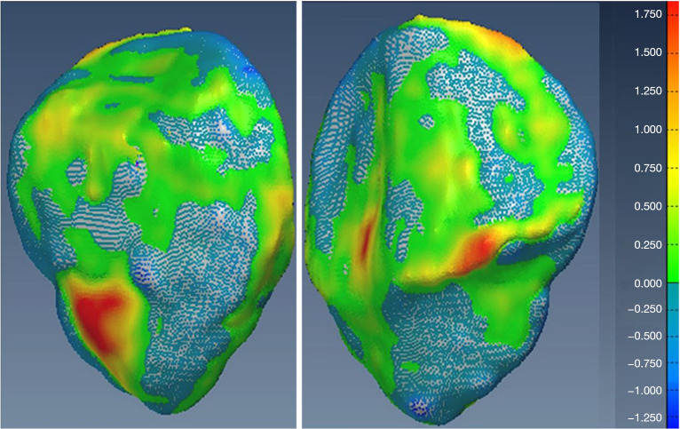
Our approach integrates the fuzzy C-means (FCM) algorithm with an adaptive region-based active contour model (ACM). Initially, the FCM algorithm assigns membership degrees to each voxel, distinguishing bone regions from surrounding soft tissues based on their likelihood of belonging to specific bone regions. Subsequently, the adaptive region-based ACM utilizes these membership degrees to guide the contour evolution and refine segmentation boundaries. To ensure clinical applicability, we further enhance our method using the marching cubes algorithm to reconstruct a three-dimensional (3D) model. We evaluated the method on six randomly selected knee joints.

RESULTS

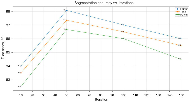
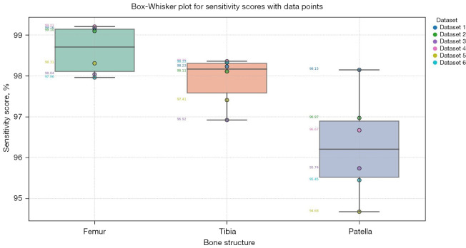
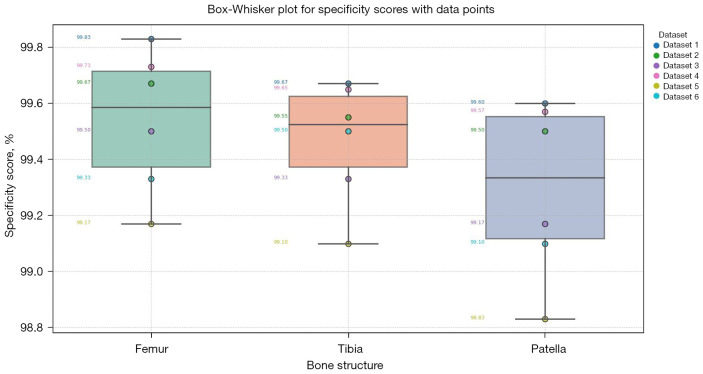
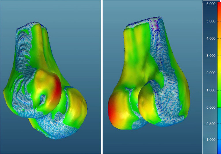
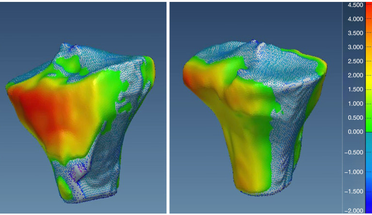
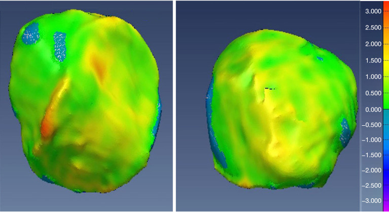
We evaluated the method using quantitative metrics such as the Dice coefficient, sensitivity, specificity, and geometrical assessment. Our method achieved high Dice scores for the femur (98.95%), tibia (98.10%), and patella (97.14%), demonstrating superior accuracy. Remarkably low root mean square distance (RSD) values were obtained for the tibia and femur (0.5±0.14 mm) and patella (0.6±0.13 mm), indicating precise segmentation.

CONCLUSIONS

The proposed method offers significant advancements in CAD systems for knee pathologies. Our approach demonstrates superior performance in achieving precise and accurate segmentation of knee bones, providing valuable insights for anatomical analysis, surgical planning, and patient-specific prostheses.

摘要

背景

准确描绘膝关节骨边界对于膝关节疾病的计算机辅助诊断(CAD)和有效的治疗规划至关重要。由于膝关节的复杂性,包括复杂的骨骼结构和重叠的软组织,当前方法在精确分割方面常常面临困难。患者解剖结构和图像质量的差异进一步加剧了这些挑战,凸显了改进技术的必要性。本文提出了一种用于从连续计算机断层扫描(CT)图像中提取膝关节骨的新型半自动分割方法。

方法

我们的方法将模糊C均值(FCM)算法与基于自适应区域的活动轮廓模型(ACM)相结合。最初,FCM算法为每个体素分配隶属度,根据其属于特定骨区域的可能性将骨区域与周围软组织区分开来。随后,基于自适应区域的ACM利用这些隶属度来引导轮廓演化并细化分割边界。为确保临床适用性,我们使用移动立方体算法进一步改进我们的方法以重建三维(3D)模型。我们在六个随机选择的膝关节上评估了该方法。

结果

我们使用诸如Dice系数、灵敏度、特异性和几何评估等定量指标评估了该方法。我们的方法在股骨(98.95%)、胫骨(98.10%)和髌骨(97.14%)上获得了较高的Dice分数,证明了卓越的准确性。胫骨、股骨(0.5±0.14毫米)和髌骨(0.6±0.13毫米)的均方根距离(RSD)值极低,表明分割精确。

结论

所提出的方法在膝关节病变的CAD系统中取得了重大进展。我们的方法在实现膝关节骨的精确和准确分割方面表现出卓越性能,为解剖分析、手术规划和定制假体提供了有价值的见解。

https://cdn.ncbi.nlm.nih.gov/pmc/blobs/0d1d/11485355/4808f2106f87/qims-14-10-7151-f26.jpg
https://cdn.ncbi.nlm.nih.gov/pmc/blobs/0d1d/11485355/e05223a75c25/qims-14-10-7151-f1.jpg
https://cdn.ncbi.nlm.nih.gov/pmc/blobs/0d1d/11485355/bde28dbfdfb8/qims-14-10-7151-f2.jpg
https://cdn.ncbi.nlm.nih.gov/pmc/blobs/0d1d/11485355/bd4cc6f572a9/qims-14-10-7151-f3.jpg
https://cdn.ncbi.nlm.nih.gov/pmc/blobs/0d1d/11485355/626cf80c85bb/qims-14-10-7151-f4.jpg
https://cdn.ncbi.nlm.nih.gov/pmc/blobs/0d1d/11485355/1d542bd9026d/qims-14-10-7151-f5.jpg
https://cdn.ncbi.nlm.nih.gov/pmc/blobs/0d1d/11485355/7c6d20aec088/qims-14-10-7151-f6.jpg
https://cdn.ncbi.nlm.nih.gov/pmc/blobs/0d1d/11485355/027463777dd9/qims-14-10-7151-f7.jpg
https://cdn.ncbi.nlm.nih.gov/pmc/blobs/0d1d/11485355/ec7e0be845ca/qims-14-10-7151-f8.jpg
https://cdn.ncbi.nlm.nih.gov/pmc/blobs/0d1d/11485355/d4ac80cb8c3d/qims-14-10-7151-f9.jpg
https://cdn.ncbi.nlm.nih.gov/pmc/blobs/0d1d/11485355/72da8af550e3/qims-14-10-7151-f10.jpg
https://cdn.ncbi.nlm.nih.gov/pmc/blobs/0d1d/11485355/f32629f563a0/qims-14-10-7151-f11.jpg
https://cdn.ncbi.nlm.nih.gov/pmc/blobs/0d1d/11485355/c939290a225e/qims-14-10-7151-f12.jpg
https://cdn.ncbi.nlm.nih.gov/pmc/blobs/0d1d/11485355/d57ffa7acb4e/qims-14-10-7151-f13.jpg
https://cdn.ncbi.nlm.nih.gov/pmc/blobs/0d1d/11485355/46fce5aeefbf/qims-14-10-7151-f14.jpg
https://cdn.ncbi.nlm.nih.gov/pmc/blobs/0d1d/11485355/215596127b9b/qims-14-10-7151-f15.jpg
https://cdn.ncbi.nlm.nih.gov/pmc/blobs/0d1d/11485355/c115f777b47e/qims-14-10-7151-f16.jpg
https://cdn.ncbi.nlm.nih.gov/pmc/blobs/0d1d/11485355/34d429dd196d/qims-14-10-7151-f17.jpg
https://cdn.ncbi.nlm.nih.gov/pmc/blobs/0d1d/11485355/31be93fd8200/qims-14-10-7151-f18.jpg
https://cdn.ncbi.nlm.nih.gov/pmc/blobs/0d1d/11485355/7188a94e248e/qims-14-10-7151-f19.jpg
https://cdn.ncbi.nlm.nih.gov/pmc/blobs/0d1d/11485355/354e5f965c99/qims-14-10-7151-f20.jpg
https://cdn.ncbi.nlm.nih.gov/pmc/blobs/0d1d/11485355/ae66c3f5508b/qims-14-10-7151-f21.jpg
https://cdn.ncbi.nlm.nih.gov/pmc/blobs/0d1d/11485355/fd6635613d94/qims-14-10-7151-f22.jpg
https://cdn.ncbi.nlm.nih.gov/pmc/blobs/0d1d/11485355/4220d4aa3db9/qims-14-10-7151-f23.jpg
https://cdn.ncbi.nlm.nih.gov/pmc/blobs/0d1d/11485355/f50909a7339b/qims-14-10-7151-f24.jpg
https://cdn.ncbi.nlm.nih.gov/pmc/blobs/0d1d/11485355/a3f27e24fbde/qims-14-10-7151-f25.jpg
https://cdn.ncbi.nlm.nih.gov/pmc/blobs/0d1d/11485355/4808f2106f87/qims-14-10-7151-f26.jpg

相似文献

1
A method framework of semi-automatic knee bone segmentation and reconstruction from computed tomography (CT) images.一种从计算机断层扫描(CT)图像中进行半自动膝关节骨分割与重建的方法框架。
Quant Imaging Med Surg. 2024 Oct 1;14(10):7151-7175. doi: 10.21037/qims-24-821. Epub 2024 Sep 26.
2
An adaptive Fuzzy C-means method utilizing neighboring information for breast tumor segmentation in ultrasound images.利用邻域信息的自适应模糊 C 均值方法在超声图像中进行乳腺肿瘤分割。
Med Phys. 2017 Jul;44(7):3752-3760. doi: 10.1002/mp.12350. Epub 2017 Jun 16.
3
Learning fuzzy clustering for SPECT/CT segmentation via convolutional neural networks.通过卷积神经网络学习用于SPECT/CT分割的模糊聚类
Med Phys. 2021 Jul;48(7):3860-3877. doi: 10.1002/mp.14903. Epub 2021 May 28.
4
Automatic segmentation of vertebral contours from CT images using fuzzy corners.使用模糊角点从CT图像中自动分割椎体轮廓。
Comput Biol Med. 2016 May 1;72:75-89. doi: 10.1016/j.compbiomed.2016.03.009. Epub 2016 Mar 18.
5
Atlas-Based Segmentation in Extraction of Knee Joint Bone Structures from CT and MR.基于图谱的 CT 和 MR 膝关节骨结构提取分割。
Sensors (Basel). 2022 Nov 19;22(22):8960. doi: 10.3390/s22228960.
6
Automatic liver segmentation by integrating fully convolutional networks into active contour models.基于全卷积网络的主动轮廓模型自动肝脏分割
Med Phys. 2019 Oct;46(10):4455-4469. doi: 10.1002/mp.13735. Epub 2019 Aug 16.
7
Applying an adaptive Otsu-based initialization algorithm to optimize active contour models for skin lesion segmentation.应用基于自适应 Otsu 的初始化算法优化用于皮肤病变分割的主动轮廓模型。
J Xray Sci Technol. 2022;30(6):1169-1184. doi: 10.3233/XST-221245.
8
Deep convolutional neural network for segmentation of knee joint anatomy.深度卷积神经网络用于膝关节解剖结构的分割。
Magn Reson Med. 2018 Dec;80(6):2759-2770. doi: 10.1002/mrm.27229. Epub 2018 May 17.
9
Fully Automatic Knee Bone Detection and Segmentation on Three-Dimensional MRI.三维磁共振成像上的全自动膝关节骨检测与分割
Diagnostics (Basel). 2022 Jan 6;12(1):123. doi: 10.3390/diagnostics12010123.
10
[Study on the accuracy of automatic segmentation of knee CT images based on deep learning].基于深度学习的膝关节CT图像自动分割准确性研究
Zhongguo Xiu Fu Chong Jian Wai Ke Za Zhi. 2022 May 15;36(5):534-539. doi: 10.7507/1002-1892.202201072.

本文引用的文献

1
Navigating the Divide: A Comprehensive Review of the Mechanical and Anatomical Axis Approaches in Total Knee Replacement.跨越分歧:全膝关节置换中机械轴和解剖轴入路的综合综述
Cureus. 2024 Apr 9;16(4):e57938. doi: 10.7759/cureus.57938. eCollection 2024 Apr.
2
Dense Multi-Scale Graph Convolutional Network for Knee Joint Cartilage Segmentation.用于膝关节软骨分割的密集多尺度图卷积网络
Bioengineering (Basel). 2024 Mar 14;11(3):278. doi: 10.3390/bioengineering11030278.
3
CartiMorph: A framework for automated knee articular cartilage morphometrics.
CartiMorph:一种用于自动膝关节软骨形态测量的框架。
Med Image Anal. 2024 Jan;91:103035. doi: 10.1016/j.media.2023.103035. Epub 2023 Nov 14.
4
Musculoskeletal CT Imaging: State-of-the-Art Advancements and Future Directions.肌肉骨骼 CT 成像:最新进展和未来方向。
Radiology. 2023 Aug;308(2):e230344. doi: 10.1148/radiol.230344.
5
Knee Bone Models From Ultrasound.膝关节超声骨模型
IEEE Trans Ultrason Ferroelectr Freq Control. 2023 Sep;70(9):1054-1063. doi: 10.1109/TUFFC.2023.3286287. Epub 2023 Aug 29.
6
Deep learning-assisted knee osteoarthritis automatic grading on plain radiographs: the value of multiview X-ray images and prior knowledge.深度学习辅助的膝关节骨关节炎X线平片自动分级:多视角X线图像及先验知识的价值
Quant Imaging Med Surg. 2023 Jun 1;13(6):3587-3601. doi: 10.21037/qims-22-1250. Epub 2023 Mar 30.
7
Magnetic resonance imaging assessments for knee segmentation and their use in combination with machine/deep learning as predictors of early osteoarthritis diagnosis and prognosis.用于膝关节分割的磁共振成像评估及其与机器学习/深度学习相结合作为早期骨关节炎诊断和预后预测指标的应用。
Ther Adv Musculoskelet Dis. 2023 Apr 28;15:1759720X231165560. doi: 10.1177/1759720X231165560. eCollection 2023.
8
A review of advances in image-guided orthopedic surgery.图像引导骨科手术进展综述。
Phys Med Biol. 2023 Jan 5;68(2). doi: 10.1088/1361-6560/acaae9.
9
Automatic femoral articular cartilage segmentation using deep learning in three-dimensional ultrasound images of the knee.在膝关节三维超声图像中利用深度学习实现股骨关节软骨的自动分割。
Osteoarthr Cartil Open. 2022 Jun 25;4(3):100290. doi: 10.1016/j.ocarto.2022.100290. eCollection 2022 Sep.
10
A novel multi-atlas segmentation approach under the semi-supervised learning framework: Application to knee cartilage segmentation.一种新的半监督学习框架下的多图谱分割方法:在膝关节软骨分割中的应用。
Comput Methods Programs Biomed. 2022 Dec;227:107208. doi: 10.1016/j.cmpb.2022.107208. Epub 2022 Oct 28.