• 文献检索
  • 文档翻译
  • 深度研究
  • 学术资讯
  • Suppr Zotero 插件Zotero 插件
  • 邀请有礼
  • 套餐&价格
  • 历史记录
应用&插件
Suppr Zotero 插件Zotero 插件浏览器插件Mac 客户端Windows 客户端微信小程序
定价
高级版会员购买积分包购买API积分包
服务
文献检索文档翻译深度研究API 文档MCP 服务
关于我们
关于 Suppr公司介绍联系我们用户协议隐私条款
关注我们

Suppr 超能文献

核心技术专利:CN118964589B侵权必究
粤ICP备2023148730 号-1Suppr @ 2026

文献检索

告别复杂PubMed语法,用中文像聊天一样搜索,搜遍4000万医学文献。AI智能推荐,让科研检索更轻松。

立即免费搜索

文件翻译

保留排版,准确专业,支持PDF/Word/PPT等文件格式,支持 12+语言互译。

免费翻译文档

深度研究

AI帮你快速写综述,25分钟生成高质量综述,智能提取关键信息,辅助科研写作。

立即免费体验

用于非典型色素性面部病变诊断的人工智能模型的比较分析

Comparative Analysis of AI Models for Atypical Pigmented Facial Lesion Diagnosis.

作者信息

Cartocci Alessandra, Luschi Alessio, Tognetti Linda, Cinotti Elisa, Farnetani Francesca, Lallas Aimilios, Paoli John, Longo Caterina, Moscarella Elvira, Tiodorovic Danica, Stanganelli Ignazio, Suppa Mariano, Dika Emi, Zalaudek Iris, Pizzichetta Maria Antonietta, Perrot Jean Luc, Cevenini Gabriele, Iadanza Ernesto, Rubegni Giovanni, Kittler Harald, Tschandl Philipp, Rubegni Pietro

机构信息

Dermatology Unit, Department of Medicine, Surgery and Neuroscience, University of Siena, 53100 Siena, Italy.

Department of Medical Biotechnologies, University of Siena, 53100 Siena, Italy.

出版信息

Bioengineering (Basel). 2024 Oct 17;11(10):1036. doi: 10.3390/bioengineering11101036.

DOI:10.3390/bioengineering11101036
PMID:39451411
原文链接:https://pmc.ncbi.nlm.nih.gov/articles/PMC11504969/
Abstract

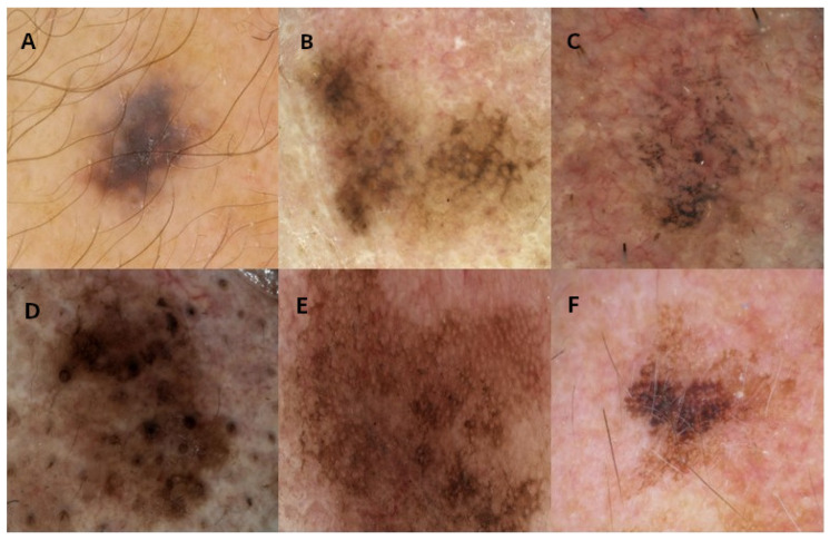
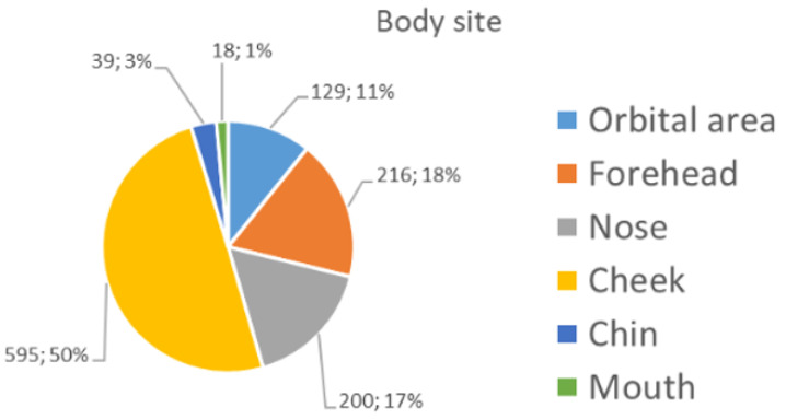
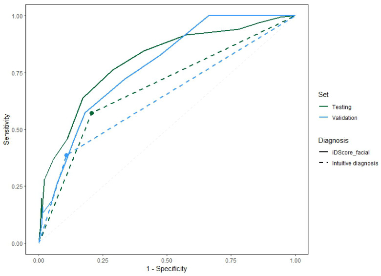
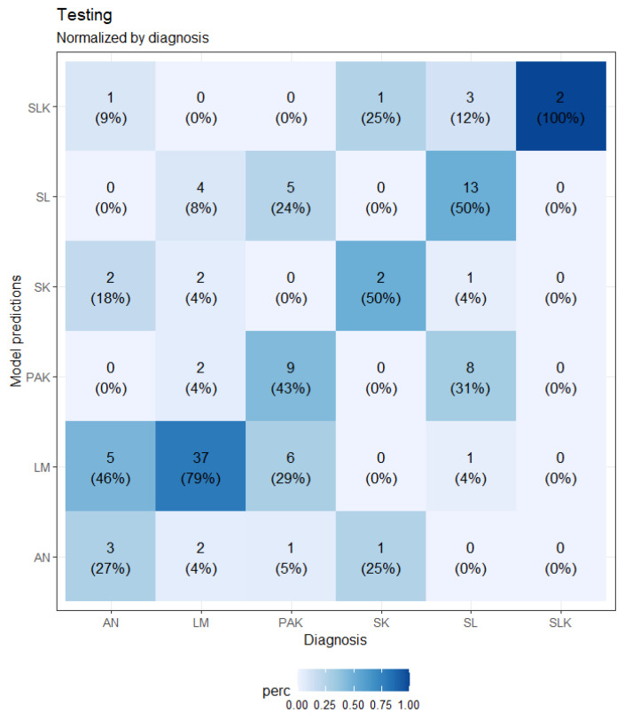
Diagnosing atypical pigmented facial lesions (aPFLs) is a challenging topic for dermatologists. Accurate diagnosis of these lesions is crucial for effective patient management, especially in dermatology, where visual assessment plays a central role. Incorrect diagnoses can result in mismanagement, delays in appropriate interventions, and potential harm. AI, however, holds the potential to enhance diagnostic accuracy and provide reliable support to clinicians. This work aimed to evaluate and compare the effectiveness of machine learning (logistic regression of lesion features and patient metadata) and deep learning (CNN analysis of images) models in dermoscopy diagnosis and the management of aPFLs. This study involved the analysis of 1197 dermoscopic images of facial lesions excised due to suspicious and histologically confirmed malignancy, classified into seven classes (lentigo maligna-LM; lentigo maligna melanoma-LMM; atypical nevi-AN; pigmented actinic keratosis-PAK; solar lentigo-SL; seborrheic keratosis-SK; and seborrheic lichenoid keratosis-SLK). Image samples were collected through the Integrated Dermoscopy Score (iDScore) project. The statistical analysis of the dataset shows that the patients mean age was 65.5 ± 14.2, and the gender was equally distributed (580 males-48.5%; 617 females-51.5%). A total of 41.7% of the sample constituted malignant lesions (LM and LMM). Meanwhile, the benign lesions were mainly PAK (19.3%), followed by SL (22.2%), AN (10.4%), SK (4.0%), and SLK (2.3%). The lesions were mainly localised in the cheek and nose areas. A stratified analysis of the assessment provided by the enrolled dermatologists was also performed, resulting in 2445 evaluations of the 1197 images (2.1 evaluations per image on average). The physicians demonstrated higher accuracy in differentiating between malignant and benign lesions (71.2%) than in distinguishing between the seven specific diagnoses across all the images (42.9%). The logistic regression model obtained a precision of 39.1%, a sensitivity of 100%, a specificity of 33.9%, and an accuracy of 53.6% on the test set, while the CNN model showed lower sensitivity (58.2%) and higher precision (47.0%), specificity (90.8%), and accuracy (59.5%) for melanoma diagnosis. This research demonstrates how AI can enhance the diagnostic accuracy in complex dermatological cases like aPFLs by integrating AI models with clinical data and evaluating different diagnostic approaches, paving the way for more precise and scalable AI applications in dermatology, showing their critical role in improving patient management and the outcomes in dermatology.

摘要

对皮肤科医生来说,诊断非典型色素沉着性面部病变(aPFLs)是一个具有挑战性的课题。准确诊断这些病变对于有效的患者管理至关重要,尤其是在皮肤科,视觉评估起着核心作用。错误的诊断可能导致管理不当、适当干预的延迟以及潜在危害。然而,人工智能有潜力提高诊断准确性并为临床医生提供可靠支持。这项工作旨在评估和比较机器学习(病变特征与患者元数据的逻辑回归)和深度学习(图像的卷积神经网络分析)模型在皮肤镜诊断和aPFLs管理中的有效性。本研究涉及对1197张因可疑且经组织学证实为恶性而切除的面部病变皮肤镜图像进行分析,这些病变分为七类(恶性雀斑样痣-LM;恶性雀斑样痣黑色素瘤-LMM;非典型痣-AN;色素性光化性角化病-PAK;日光性雀斑样痣-SL;脂溢性角化病-SK;以及脂溢性苔藓样角化病-SLK)。图像样本通过综合皮肤镜评分(iDScore)项目收集。数据集的统计分析表明,患者的平均年龄为6岁5.5±14.2岁,性别分布均衡(580名男性-48.5%;617名女性-51.5%)。样本中共有41.7%为恶性病变(LM和LMM)。同时,良性病变主要为PAK(19.3%),其次是SL(22.2%)、AN(10.4%)、SK(4.0%)和SLK(2.3%)。病变主要位于脸颊和鼻子区域。还对参与研究的皮肤科医生提供的评估进行了分层分析,对1197张图像进行了2445次评估(平均每张图像2.1次评估)。医生在区分恶性和良性病变方面(71.2%)比在区分所有图像中的七种具体诊断方面(42.9%)表现出更高的准确性。逻辑回归模型在测试集上的精确率为39.1%,灵敏度为100%,特异度为33.9%,准确率为53.6%,而卷积神经网络模型在黑色素瘤诊断方面显示出较低的灵敏度(58.2%)和较高的精确率(47.0%)、特异度(90.8%)和准确率(59.5%)。这项研究展示了人工智能如何通过将人工智能模型与临床数据相结合并评估不同的诊断方法,提高aPFLs等复杂皮肤病病例的诊断准确性,为皮肤科更精确、可扩展的人工智能应用铺平道路,显示出它们在改善皮肤科患者管理和治疗结果方面的关键作用。

https://cdn.ncbi.nlm.nih.gov/pmc/blobs/a127/11504969/f2be984c1624/bioengineering-11-01036-g007.jpg
https://cdn.ncbi.nlm.nih.gov/pmc/blobs/a127/11504969/94bb277683c7/bioengineering-11-01036-g001.jpg
https://cdn.ncbi.nlm.nih.gov/pmc/blobs/a127/11504969/ecd0aa4b37d3/bioengineering-11-01036-g002.jpg
https://cdn.ncbi.nlm.nih.gov/pmc/blobs/a127/11504969/e59c3a534426/bioengineering-11-01036-g003.jpg
https://cdn.ncbi.nlm.nih.gov/pmc/blobs/a127/11504969/d709992c1c30/bioengineering-11-01036-g004.jpg
https://cdn.ncbi.nlm.nih.gov/pmc/blobs/a127/11504969/5928a6311091/bioengineering-11-01036-g005.jpg
https://cdn.ncbi.nlm.nih.gov/pmc/blobs/a127/11504969/fa83c9146211/bioengineering-11-01036-g006.jpg
https://cdn.ncbi.nlm.nih.gov/pmc/blobs/a127/11504969/f2be984c1624/bioengineering-11-01036-g007.jpg

相似文献

1
Comparative Analysis of AI Models for Atypical Pigmented Facial Lesion Diagnosis.用于非典型色素性面部病变诊断的人工智能模型的比较分析
Bioengineering (Basel). 2024 Oct 17;11(10):1036. doi: 10.3390/bioengineering11101036.
2
Dermoscopy of atypical pigmented lesions of the face: Variation according to facial areas.面部非典型色素性皮损的皮肤镜检查:与面部区域的变化有关。
Exp Dermatol. 2023 Dec;32(12):2166-2172. doi: 10.1111/exd.14941. Epub 2023 Sep 28.
3
A risk-scoring model for the differential diagnosis of lentigo maligna and other atypical pigmented facial lesions of the face: The facial iDScore.一种用于鉴别诊断良性色素痣和其他面部非典型色素性病变的风险评分模型:面部 iDScore。
J Eur Acad Dermatol Venereol. 2023 Nov;37(11):2301-2310. doi: 10.1111/jdv.19360. Epub 2023 Jul 26.
4
Role of In Vivo Reflectance Confocal Microscopy in the Analysis of Melanocytic Lesions.体内反射共聚焦显微镜在黑素细胞性病变分析中的作用
Acta Dermatovenerol Croat. 2018 Apr;26(1):64-67.
5
Development and Implementation of a Web-Based International Registry Dedicated to Atypical Pigmented Skin Lesions of the Face: Teledermatologic Investigation on Epidemiology and Risk Factors.基于网络的面部非典型色素性皮肤病变国际登记处的开发与实施:皮肤疾病流行病学和风险因素的远程皮肤病学调查
Telemed J E Health. 2023 Sep;29(9):1356-1365. doi: 10.1089/tmj.2022.0456. Epub 2023 Feb 8.
6
A comparison of dermoscopic features among lentigo senilis/initial seborrheic keratosis, seborrheic keratosis, lentigo maligna and lentigo maligna melanoma on the face.面部脂溢性角化病/早期脂溢性角化病、脂溢性角化病、恶性雀斑样痣及恶性雀斑样痣黑色素瘤的皮肤镜特征比较
J Dermatol. 2004 Nov;31(11):884-9. doi: 10.1111/j.1346-8138.2004.tb00621.x.
7
Fluorescence-advanced videodermatoscopy (FAV) for the differential diagnosis of suspicious facial lesions: A single-centre experience with pattern analysis and histopathological correlation.荧光增强视频皮肤镜(FAV)在可疑面部病变中的鉴别诊断:基于模式分析和组织病理学相关性的单中心经验。
Photodermatol Photoimmunol Photomed. 2022 May;38(3):266-276. doi: 10.1111/phpp.12748. Epub 2021 Nov 3.
8
Comparison of Ophthalmologists versus Dermatologists for the Diagnosis and Management of Periorbital Atypical Pigmented Skin Lesions.眼科医生与皮肤科医生对眶周非典型色素沉着性皮肤病变的诊断与管理比较
J Clin Med. 2024 Aug 14;13(16):4787. doi: 10.3390/jcm13164787.
9
The dermoscopic inverse approach significantly improves the accuracy of human readers for lentigo maligna diagnosis.皮肤镜下反推法显著提高了人眼阅读者对恶性雀斑样痣诊断的准确性。
J Am Acad Dermatol. 2021 Feb;84(2):381-389. doi: 10.1016/j.jaad.2020.06.085. Epub 2020 Jun 24.
10
Dermoscopic Differentiation of Facial Lentigo Maligna from Pigmented Actinic Keratosis and Solar Lentigines.面部恶性雀斑样痣与色素性日光性角化病及日光性雀斑的皮肤镜鉴别
Acta Dermatovenerol Croat. 2019 Sep;27(3):146-152.

引用本文的文献

1
Deep Learning for Melanoma Detection: A Deep Learning Approach to Differentiating Malignant Melanoma from Benign Melanocytic Nevi.用于黑色素瘤检测的深度学习:一种区分恶性黑色素瘤与良性黑素细胞痣的深度学习方法。
Cancers (Basel). 2024 Dec 25;17(1):28. doi: 10.3390/cancers17010028.

本文引用的文献

1
The Arrival of Artificial Intelligence Large Language Models and Vision-Language Models: A Potential to Possible Change in the Paradigm of Healthcare Delivery in Dermatology.人工智能大语言模型和视觉语言模型的到来:皮肤病学医疗服务模式可能发生变革的潜力。
J Invest Dermatol. 2024 Jun;144(6):1186-1188. doi: 10.1016/j.jid.2023.10.046. Epub 2024 Feb 1.
2
Overdiagnosis in malignant melanoma: a scoping review.恶性黑色素瘤的过度诊断:范围综述。
BMJ Evid Based Med. 2024 Jan 19;29(1):17-28. doi: 10.1136/bmjebm-2023-112341.
3
SBXception: A Shallower and Broader Xception Architecture for Efficient Classification of Skin Lesions.
SBXception:一种用于皮肤病变高效分类的更浅且更宽的Xception架构。
Cancers (Basel). 2023 Jul 13;15(14):3604. doi: 10.3390/cancers15143604.
4
A risk-scoring model for the differential diagnosis of lentigo maligna and other atypical pigmented facial lesions of the face: The facial iDScore.一种用于鉴别诊断良性色素痣和其他面部非典型色素性病变的风险评分模型:面部 iDScore。
J Eur Acad Dermatol Venereol. 2023 Nov;37(11):2301-2310. doi: 10.1111/jdv.19360. Epub 2023 Jul 26.
5
Artificial Intelligence in Dermatology Image Analysis: Current Developments and Future Trends.皮肤病学图像分析中的人工智能:当前进展与未来趋势。
J Clin Med. 2022 Nov 18;11(22):6826. doi: 10.3390/jcm11226826.
6
An Updated Algorithm Integrated With Patient Data for the Differentiation of Atypical Nevi From Early Melanomas: the idScore 2021.一种结合患者数据用于鉴别非典型痣与早期黑色素瘤的更新算法:idScore 2021
Dermatol Pract Concept. 2022 Jul 1;12(3):e2022134. doi: 10.5826/dpc.1203a134. eCollection 2022 Jul.
7
Global Burden of Cutaneous Melanoma in 2020 and Projections to 2040.2020 年全球皮肤黑色素瘤负担及 2040 年预测。
JAMA Dermatol. 2022 May 1;158(5):495-503. doi: 10.1001/jamadermatol.2022.0160.
8
Assessment of Diagnostic Accuracy of Dermoscopic Structures and Patterns Used in Melanoma Detection: A Systematic Review and Meta-analysis.评估用于黑色素瘤检测的皮肤镜结构和模式的诊断准确性:系统评价和荟萃分析。
JAMA Dermatol. 2021 Sep 1;157(9):1078-1088. doi: 10.1001/jamadermatol.2021.2845.
9
A new deep learning approach integrated with clinical data for the dermoscopic differentiation of early melanomas from atypical nevi.一种新的深度学习方法与临床数据相结合,用于对早期黑色素瘤与非典型痣进行皮肤镜鉴别。
J Dermatol Sci. 2021 Feb;101(2):115-122. doi: 10.1016/j.jdermsci.2020.11.009. Epub 2020 Dec 2.
10
Multimodal skin lesion classification using deep learning.基于深度学习的多模态皮肤损伤分类。
Exp Dermatol. 2018 Nov;27(11):1261-1267. doi: 10.1111/exd.13777. Epub 2018 Sep 27.