Al Jada Iyad, Jabareen Maaweya, Alhroub Wasef, Oweidat Majd
Department of Surgery, Hebron University, Hebron, Palestine.
Faculty of Medicine, Hebron University, Hebron, Palestine.
Int J Surg Case Rep. 2024 Dec;125:110504. doi: 10.1016/j.ijscr.2024.110504. Epub 2024 Oct 23.
Mature cystic teratomas of the pancreas, also known as dermoid cysts, are exceptionally rare tumors characterized by well-differentiated parenchymal tissues. Typically containing diverse tissues from all three germ layers, these teratomas are most commonly found in the ovaries and testes, with infrequent occurrences in the pancreas.
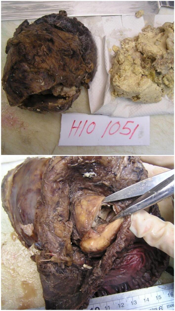
A 30-year-old male with type 2 diabetes mellitus presented with elevated liver enzymes and serum CEA levels. A CT scan detected an 8.8 × 7.2 cm retroperitoneal mass with calcifications. Due to the tumor's involvement with critical structures, a Whipple procedure was performed. Post-surgery, the tumor was confirmed to be a mature cystic teratoma, and the patient experienced a smooth recovery.
Pancreatic teratomas are rare, typically affecting younger patients and predominantly occurring in the body or head of the pancreas. These tumors, often categorized into mature and immature types. Diagnosis relies on imaging techniques such as ultrasound, CT, and MRI, which reveal key features like fat, calcifications, and fat-fluid levels. Differential diagnoses include various pancreatic cystic lesions. Surgical resection is the primary treatment, and this case highlights the diagnostic challenges and the critical role of imaging in guiding surgical decisions.
This report describes a rare case of a pancreatic mature cystic teratoma, one of only 52 documented cases. Despite the absence of significant symptoms, imaging revealed a large mass, and Whipple procedure was performed due to its complex relationship with vital structures. This case illustrates the diagnostic and therapeutic challenges associated with such rare tumors.
胰腺成熟囊性畸胎瘤,也称为皮样囊肿,是极为罕见的肿瘤,其特征为实质组织分化良好。这些畸胎瘤通常包含来自所有三个胚层的不同组织,最常见于卵巢和睾丸,在胰腺中很少见。
一名30岁的2型糖尿病男性患者出现肝酶和血清癌胚抗原水平升高。CT扫描发现一个8.8×7.2cm的腹膜后肿块伴有钙化。由于肿瘤累及关键结构,遂进行了惠普尔手术。术后,肿瘤被确诊为成熟囊性畸胎瘤,患者恢复顺利。
胰腺畸胎瘤罕见,通常影响年轻患者,主要发生在胰腺体部或头部。这些肿瘤常分为成熟型和未成熟型。诊断依赖于超声、CT和MRI等成像技术,这些技术可显示脂肪、钙化和脂液平面等关键特征。鉴别诊断包括各种胰腺囊性病变。手术切除是主要治疗方法,本病例突出了诊断挑战以及成像在指导手术决策中的关键作用。
本报告描述了一例罕见的胰腺成熟囊性畸胎瘤病例,是仅有的52例有记录的病例之一。尽管没有明显症状,但成像显示有一个大肿块,由于其与重要结构的复杂关系,进行了惠普尔手术。本病例说明了与此类罕见肿瘤相关的诊断和治疗挑战。