• 文献检索
  • 文档翻译
  • 深度研究
  • 学术资讯
  • Suppr Zotero 插件Zotero 插件
  • 邀请有礼
  • 套餐&价格
  • 历史记录
应用&插件
Suppr Zotero 插件Zotero 插件浏览器插件Mac 客户端Windows 客户端微信小程序
定价
高级版会员购买积分包购买API积分包
服务
文献检索文档翻译深度研究API 文档MCP 服务
关于我们
关于 Suppr公司介绍联系我们用户协议隐私条款
关注我们

Suppr 超能文献

核心技术专利:CN118964589B侵权必究
粤ICP备2023148730 号-1Suppr @ 2026

文献检索

告别复杂PubMed语法,用中文像聊天一样搜索,搜遍4000万医学文献。AI智能推荐,让科研检索更轻松。

立即免费搜索

文件翻译

保留排版,准确专业,支持PDF/Word/PPT等文件格式,支持 12+语言互译。

免费翻译文档

深度研究

AI帮你快速写综述,25分钟生成高质量综述,智能提取关键信息,辅助科研写作。

立即免费体验

利用深度学习分析对切除的结直肠癌肝转移进行组织病理学生长模式分类。

Classifying histopathological growth patterns for resected colorectal liver metastasis with a deep learning analysis.

机构信息

Department of Surgical Oncology and Gastrointestinal Surgery, Erasmus MC Cancer Institute, University Medical Center Rotterdam, Rotterdam, The Netherlands.

Departments of Pathology, Radboud University Medical Center, Nijmegen, The Netherlands.

出版信息

BJS Open. 2024 Oct 29;8(6). doi: 10.1093/bjsopen/zrae127.

DOI:10.1093/bjsopen/zrae127
PMID:39471410
原文链接:https://pmc.ncbi.nlm.nih.gov/articles/PMC11523050/
Abstract

BACKGROUND

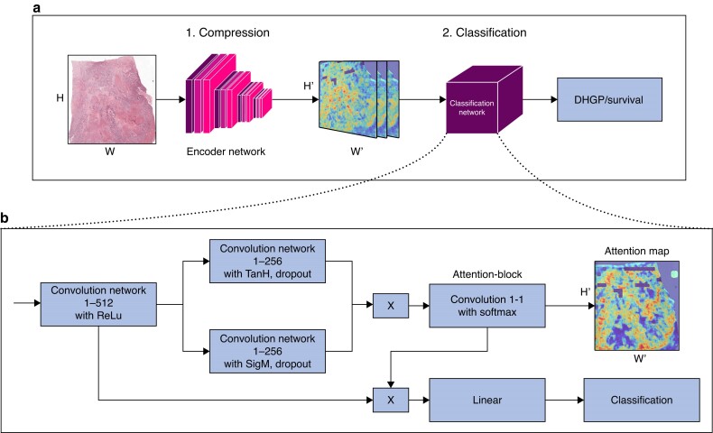
Histopathological growth patterns are one of the strongest prognostic factors in patients with resected colorectal liver metastases. Development of an efficient, objective and ideally automated histopathological growth pattern scoring method can substantially help the implementation of histopathological growth pattern assessment in daily practice and research. This study aimed to develop and validate a deep-learning algorithm, namely neural image compression, to distinguish desmoplastic from non-desmoplastic histopathological growth patterns of colorectal liver metastases based on digital haematoxylin and eosin-stained slides.

METHODS

The algorithm was developed using digitalized whole-slide images obtained in a single-centre (Erasmus MC Cancer Institute, the Netherlands) cohort of patients who underwent first curative intent resection for colorectal liver metastases between January 2000 and February 2019. External validation was performed on whole-slide images of patients resected between October 2004 and December 2017 in another institution (Radboud University Medical Center, the Netherlands). The outcomes of interest were the automated classification of dichotomous hepatic growth patterns, distinguishing between desmoplastic hepatic growth pattern and non-desmoplatic growth pattern by a deep-learning model; secondary outcome was the correlation of these classifications with overall survival in the histopathology manual-assessed histopathological growth pattern and those assessed using neural image compression.

RESULTS

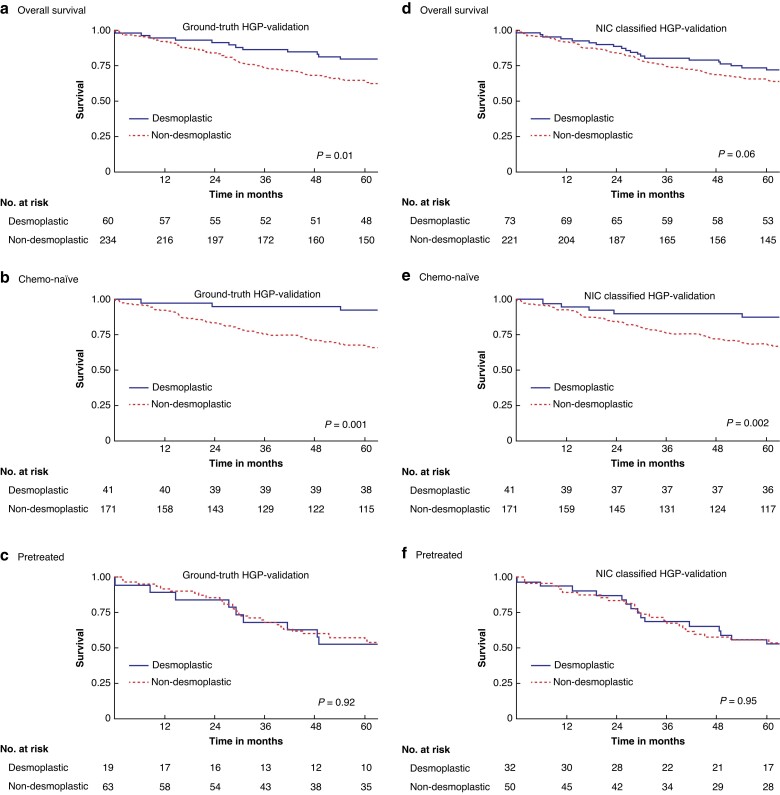
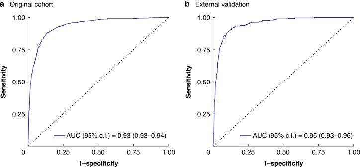
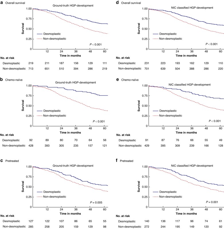
Nine hundred and thirty-two patients, corresponding to 3.641 whole-slide images, were reviewed to develop the algorithm and 870 whole-slide images were used for external validation. Median follow-up for the development and the validation cohorts was 43 and 29 months respectively. The neural image compression approach achieved significant discriminatory power to classify 100% desmoplastic histopathological growth pattern with an area under the curve of 0.93 in the development cohort and 0.95 upon external validation. Both the histopathology manual-scored histopathological growth pattern and neural image compression-classified histopathological growth pattern achieved a similar multivariable hazard ratio for desmoplastic versus non-desmoplastic growth pattern in the development cohort (histopathology manual score: 0.63 versus neural image compression: 0.64) and in the validation cohort (histopathology manual score: 0.40 versus neural image compression: 0.48).

CONCLUSIONS

The neural image compression approach is suitable for pathology-based classification tasks of colorectal liver metastases.

摘要

背景

在接受结直肠肝转移切除术的患者中,组织病理学生长模式是最强的预后因素之一。开发一种高效、客观且理想的自动化组织病理学生长模式评分方法,可以极大地帮助在日常实践和研究中实施组织病理学生长模式评估。本研究旨在开发和验证一种深度学习算法,即神经图像压缩,以区分结直肠肝转移的纤维组织增生型和非纤维组织增生型组织病理学生长模式,基于数字化苏木精和伊红染色切片。

方法

该算法是使用 2000 年 1 月至 2019 年 2 月期间在一家单中心(荷兰伊拉斯谟医学中心)接受首次治愈性意向切除术治疗的患者的数字化全切片图像开发的。外部验证是在另一家机构(荷兰拉德堡德大学医学中心)于 2004 年 10 月至 2017 年 12 月期间切除的患者的全切片图像上进行的。主要结局是通过深度学习模型对二分类肝生长模式进行自动分类,区分纤维组织增生型肝生长模式和非纤维组织增生型生长模式;次要结局是这些分类与组织病理学手动评估的组织病理学生长模式以及使用神经图像压缩评估的组织病理学生长模式的总体生存率之间的相关性。

结果

共回顾了 932 例患者,对应 3641 张全切片图像,用于开发算法,870 张全切片图像用于外部验证。发展队列和验证队列的中位随访时间分别为 43 个月和 29 个月。神经图像压缩方法在开发队列中达到了显著的区分能力,以 100%的准确率对 100%的纤维组织增生型组织病理学生长模式进行分类,曲线下面积为 0.93,在外部验证中达到了 0.95。组织病理学手动评分的组织病理学生长模式和神经图像压缩分类的组织病理学生长模式在发展队列(组织病理学手动评分:0.63 与神经图像压缩:0.64)和验证队列(组织病理学手动评分:0.40 与神经图像压缩:0.48)中均具有类似的多变量风险比,用于纤维组织增生型与非纤维组织增生型生长模式的比较。

结论

神经图像压缩方法适用于结直肠肝转移的基于病理学的分类任务。

https://cdn.ncbi.nlm.nih.gov/pmc/blobs/6afc/11523050/f949f8442592/zrae127f4.jpg
https://cdn.ncbi.nlm.nih.gov/pmc/blobs/6afc/11523050/e4946b894407/zrae127f1.jpg
https://cdn.ncbi.nlm.nih.gov/pmc/blobs/6afc/11523050/e4496e648aba/zrae127f2.jpg
https://cdn.ncbi.nlm.nih.gov/pmc/blobs/6afc/11523050/efa8adf79ad4/zrae127f3.jpg
https://cdn.ncbi.nlm.nih.gov/pmc/blobs/6afc/11523050/f949f8442592/zrae127f4.jpg

相似文献

1
Classifying histopathological growth patterns for resected colorectal liver metastasis with a deep learning analysis.利用深度学习分析对切除的结直肠癌肝转移进行组织病理学生长模式分类。
BJS Open. 2024 Oct 29;8(6). doi: 10.1093/bjsopen/zrae127.
2
Histopathological growth patterns as biomarker for adjuvant systemic chemotherapy in patients with resected colorectal liver metastases.切除术后结直肠癌肝转移患者的组织病理学生长模式作为辅助全身化疗的生物标志物。
Clin Exp Metastasis. 2020 Oct;37(5):593-605. doi: 10.1007/s10585-020-10048-w. Epub 2020 Jul 20.
3
Histopathological growth patterns of resected non-colorectal, non-neuroendocrine liver metastases: a retrospective multicenter study.切除的非结直肠、非神经内分泌肝转移瘤的组织病理学生长模式:一项回顾性多中心研究。
Clin Exp Metastasis. 2022 Jun;39(3):433-442. doi: 10.1007/s10585-022-10153-y. Epub 2022 Feb 6.
4
Histopathological Growth Patterns and Survival After Resection of Colorectal Liver Metastasis: An External Validation Study.结直肠肝转移切除术后的组织病理学生长模式和生存:一项外部验证研究。
JNCI Cancer Spectr. 2021 Mar 21;5(3). doi: 10.1093/jncics/pkab026. eCollection 2021 Jun.
5
Preoperative systemic chemotherapy alters the histopathological growth patterns of colorectal liver metastases.术前全身化疗改变结直肠癌肝转移的组织病理学生长模式。
J Pathol Clin Res. 2022 Jan;8(1):48-64. doi: 10.1002/cjp2.235. Epub 2021 Sep 4.
6
Angiogenic desmoplastic histopathological growth pattern as a prognostic marker of good outcome in patients with colorectal liver metastases.结直肠肝转移患者中血管生成性促结缔组织增生型组织病理学生长模式作为预后良好的标志物。
Angiogenesis. 2019 May;22(2):355-368. doi: 10.1007/s10456-019-09661-5. Epub 2019 Jan 12.
7
Automated curation of large-scale cancer histopathology image datasets using deep learning.利用深度学习对大规模癌症组织病理学图像数据集进行自动化注释。
Histopathology. 2024 Jun;84(7):1139-1153. doi: 10.1111/his.15159. Epub 2024 Feb 26.
8
Replacement and desmoplastic histopathological growth patterns in cutaneous melanoma liver metastases: frequency, characteristics, and robust prognostic value.皮肤黑色素瘤肝转移中的替代型和促纤维组织增生型组织病理学生长模式:发生率、特征及可靠的预后价值。
J Pathol Clin Res. 2020 Jul;6(3):195-206. doi: 10.1002/cjp2.161. Epub 2020 Apr 18.
9
The relationship between primary colorectal cancer histology and the histopathological growth patterns of corresponding liver metastases.原发性结直肠癌组织学与相应肝转移的组织病理学生长模式之间的关系。
BMC Cancer. 2022 Aug 22;22(1):911. doi: 10.1186/s12885-022-09994-3.
10
Association between primary tumor characteristics and histopathological growth pattern of liver metastases in colorectal cancer.结直肠癌中原发性肿瘤特征与肝转移灶组织病理学生长模式之间的关联
Clin Exp Metastasis. 2023 Oct;40(5):431-440. doi: 10.1007/s10585-023-10221-x. Epub 2023 Jul 15.

引用本文的文献

1
The prognostic significance of histopathological growth patterns in liver metastases undergoing surgery: a systematic review and meta-analysis.接受手术的肝转移瘤组织病理学生长模式的预后意义:一项系统评价和荟萃分析。
Clin Exp Metastasis. 2025 Jul 15;42(5):41. doi: 10.1007/s10585-025-10361-2.
2
Advances and challenges in pathomics for liver cancer: From diagnosis to prognostic stratification.肝癌病理组学的进展与挑战:从诊断到预后分层
World J Clin Oncol. 2025 Jun 24;16(6):107646. doi: 10.5306/wjco.v16.i6.107646.
3
Factors affecting survival prognosis of patients with rectal cancer after neoadjuvant chemoradiotherapy.

本文引用的文献

1
An idiosyncratic zonated stroma encapsulates desmoplastic liver metastases and originates from injured liver.独特的区域性基质包绕着促纤维增生性肝转移瘤,并起源于受损的肝脏。
Nat Commun. 2023 Aug 18;14(1):5024. doi: 10.1038/s41467-023-40688-x.
2
Histopathological Growth Pattern in Colorectal Liver Metastasis and The Tumor Immune Microenvironment.结直肠癌肝转移的组织病理学生长模式与肿瘤免疫微环境
Cancers (Basel). 2022 Dec 28;15(1):181. doi: 10.3390/cancers15010181.
3
Histopathological growth patterns of liver metastasis: updated consensus guidelines for pattern scoring, perspectives and recent mechanistic insights.
新辅助放化疗后直肠癌患者生存预后的影响因素
Front Oncol. 2025 May 15;15:1562634. doi: 10.3389/fonc.2025.1562634. eCollection 2025.
肝脏转移瘤的组织病理学生长模式:更新的共识指南用于模式评分、观点和最近的机制见解。
Br J Cancer. 2022 Oct;127(6):988-1013. doi: 10.1038/s41416-022-01859-7. Epub 2022 Jun 1.
4
Predicting 10-year survival after resection of colorectal liver metastases; an international study including biomarkers and perioperative treatment.预测结直肠肝转移切除术后 10 年生存率:一项包含生物标志物和围手术期治疗的国际研究。
Eur J Cancer. 2022 Jun;168:25-33. doi: 10.1016/j.ejca.2022.01.012. Epub 2022 Apr 14.
5
Treatment of metachronous colorectal cancer metastases in the Netherlands: A population-based study.荷兰异时性结直肠癌转移的治疗:一项基于人群的研究。
Eur J Surg Oncol. 2022 May;48(5):1104-1109. doi: 10.1016/j.ejso.2021.12.004. Epub 2021 Dec 4.
6
Histopathological Growth Patterns and Survival After Resection of Colorectal Liver Metastasis: An External Validation Study.结直肠肝转移切除术后的组织病理学生长模式和生存:一项外部验证研究。
JNCI Cancer Spectr. 2021 Mar 21;5(3). doi: 10.1093/jncics/pkab026. eCollection 2021 Jun.
7
Deep learning in histopathology: the path to the clinic.深度学习在组织病理学中的应用:通往临床的道路。
Nat Med. 2021 May;27(5):775-784. doi: 10.1038/s41591-021-01343-4. Epub 2021 May 14.
8
Data-efficient and weakly supervised computational pathology on whole-slide images.基于全切片图像的数据高效和弱监督计算病理学。
Nat Biomed Eng. 2021 Jun;5(6):555-570. doi: 10.1038/s41551-020-00682-w. Epub 2021 Mar 1.
9
Designing deep learning studies in cancer diagnostics.设计癌症诊断的深度学习研究。
Nat Rev Cancer. 2021 Mar;21(3):199-211. doi: 10.1038/s41568-020-00327-9. Epub 2021 Jan 29.
10
Deep learning in cancer pathology: a new generation of clinical biomarkers.深度学习在癌症病理学中的应用:新一代临床生物标志物。
Br J Cancer. 2021 Feb;124(4):686-696. doi: 10.1038/s41416-020-01122-x. Epub 2020 Nov 18.