CNRS, LMR, UMR 9008, Université de Reims Champagne Ardenne, Reims, France.
CRESTIC, Université de Reims Champagne Ardenne, Reims, France.
PLoS One. 2024 Nov 1;19(11):e0312822. doi: 10.1371/journal.pone.0312822. eCollection 2024.
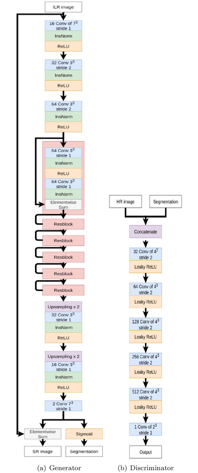
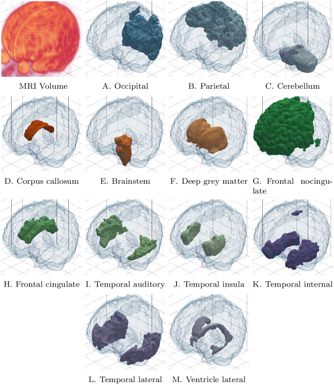
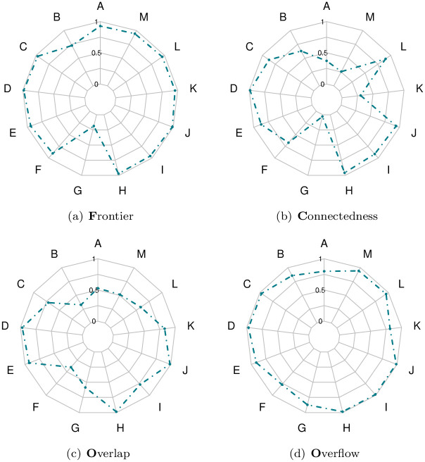
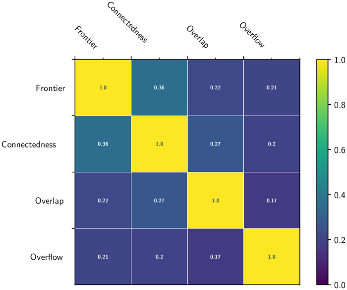
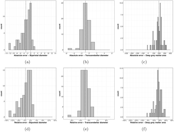
Magnetic resonance imaging (MRI) is a powerful tool for observing and assessing the properties of brain tissue and structures. In particular, in the context of neonatal care, MR images can be used to analyze neurodevelopmental problems that may arise in premature newborns. However, the intrinsic properties of newborn MR images, combined with the high variability of MR acquisition in a clinical setting, result in complex and heterogeneous images. Segmentation methods dedicated to the processing of clinical data are essential for obtaining relevant biomarkers. In this context, the design of quality control protocols for the associated segmentation is a cornerstone for guaranteeing the accuracy and usefulness of these inferred biomarkers. In recent work, we have proposed a new method, SegSRGAN, designed for super-resolution reconstruction and segmentation of specific brain structures. In this article, we first propose an extension of SegSRGAN from binary segmentation to multi-label segmentation, leading then to a partitioning of an MR image into several labels, each corresponding to a specific brain tissue/area. Secondly, we propose a segmentation quality control protocol designed to assess the performance of the proposed method with regard to this specific parcellation task in neonatal MR imaging. In particular, we combine scores derived from expert analysis, morphometric measurements and topological properties of the structures studied. This segmentation quality control can enable clinicians to select reliable segmentations for clinical analysis, starting with correlations between perinatal risk factors, regional volumes and specific dimensions of cognitive development. Based on this protocol, we are investigating the strengths and weaknesses of SegSRGAN and its potential suitability for clinical research in the context of morphometric analysis of brain structure in preterm infants, and to potentially design new biomarkers of neurodevelopment. The proposed study focuses on MR images from the EPIRMEX dataset, collected as part of a national cohort study. In particular, this work represents a first step towards the design of 3-dimensional neonatal brain morphometry based on segmentation. The (free and open-source) code of multilabel SegSRGAN is publicly available at the following URL: https://doi.org/10.5281/zenodo.12659424.
磁共振成像(MRI)是观察和评估脑组织和结构特性的强大工具。特别是在新生儿护理领域,MR 图像可用于分析早产儿可能出现的神经发育问题。然而,新生儿 MR 图像的固有特性,加上临床环境中 MRI 采集的高度可变性,导致图像复杂且具有异质性。专门用于处理临床数据的分割方法对于获得相关生物标志物至关重要。在这种情况下,相关分割的质量控制协议的设计是保证这些推断出的生物标志物准确性和有用性的基石。在最近的工作中,我们提出了一种新的方法 SegSRGAN,用于特定脑结构的超分辨率重建和分割。在本文中,我们首先将 SegSRGAN 从二进制分割扩展到多标签分割,从而将 MR 图像分割成几个标签,每个标签对应于特定的脑组织/区域。其次,我们提出了一种分割质量控制协议,旨在评估所提出的方法在新生儿 MR 成像中特定分割任务中的性能。特别是,我们结合了来自专家分析、形态测量和所研究结构拓扑特性的评分。这种分割质量控制可以使临床医生能够选择可靠的分割进行临床分析,首先是围产期风险因素、区域体积和认知发展特定维度之间的相关性。基于该协议,我们正在研究 SegSRGAN 的优缺点及其在早产儿脑结构形态测量分析中的临床研究中的潜在适用性,以及设计神经发育新生物标志物的潜力。本研究侧重于 EPIRMEX 数据集的 MR 图像,这些图像是作为国家队列研究的一部分收集的。特别是,这项工作代表了基于分割设计三维新生儿脑形态测量的第一步。多标签 SegSRGAN 的(免费和开源)代码可在以下 URL 获得:https://doi.org/10.5281/zenodo.12659424。