Guo Ting, Winterburn Julie L, Pipitone Jon, Duerden Emma G, Park Min Tae M, Chau Vann, Poskitt Kenneth J, Grunau Ruth E, Synnes Anne, Miller Steven P, Mallar Chakravarty M
Neurosciences and Mental Health, The Hospital for Sick Children Research Institute, Toronto, ON, Canada; Department of Paediatrics, The Hospital for Sick Children and the University of Toronto, Toronto, ON, Canada.
Institute of Biomedical Engineering, University of Toronto, Toronto, ON, Canada; Kimel Family Translational Imaging, Genetics Research Laboratory, Research Imaging Centre, Centre for Addiction and Mental Health, Toronto, Canada.
Neuroimage Clin. 2015 Aug 24;9:176-93. doi: 10.1016/j.nicl.2015.07.019. eCollection 2015.
The hippocampus, a medial temporal lobe structure central to learning and memory, is particularly vulnerable in preterm-born neonates. To date, segmentation of the hippocampus for preterm-born neonates has not yet been performed early-in-life (shortly after birth when clinically stable). The present study focuses on the development and validation of an automatic segmentation protocol that is based on the MAGeT-Brain (Multiple Automatically Generated Templates) algorithm to delineate the hippocampi of preterm neonates on their brain MRIs acquired at not only term-equivalent age but also early-in-life.
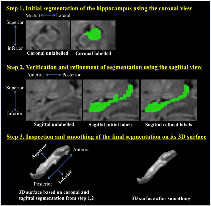
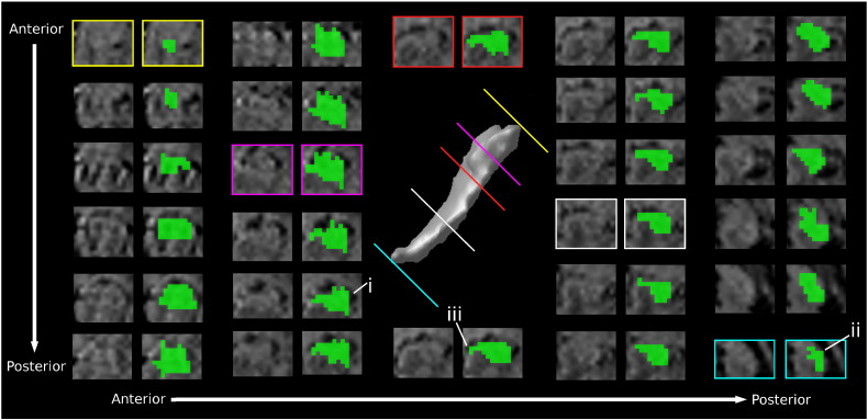
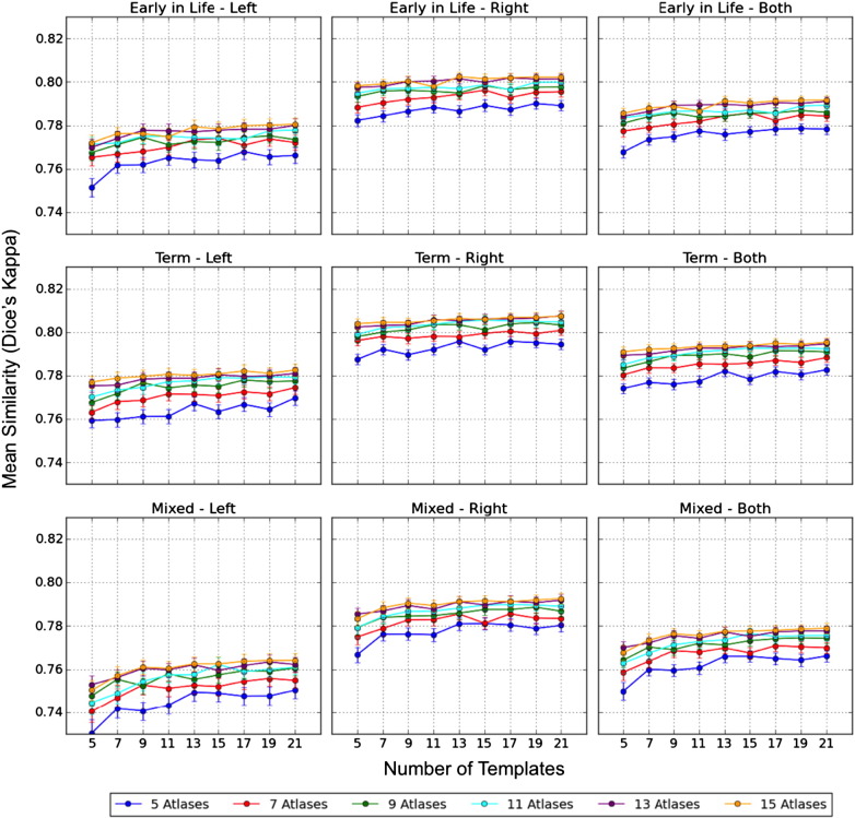
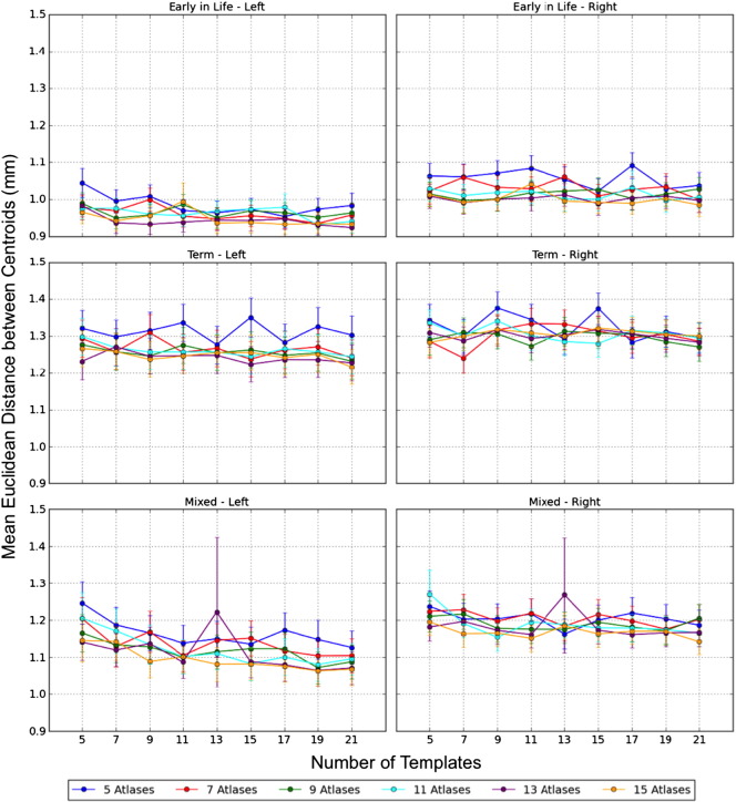
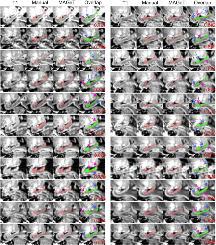
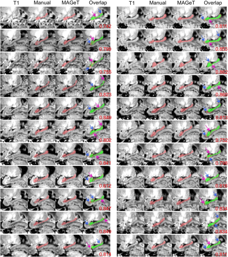
First, we present a three-step manual segmentation protocol to delineate the hippocampus for preterm neonates and apply this protocol on 22 early-in-life and 22 term images. These manual segmentations are considered the gold standard in assessing the automatic segmentations. MAGeT-Brain, automatic hippocampal segmentation pipeline, requires only a small number of input atlases and reduces the registration and resampling errors by employing an intermediate template library. We assess the segmentation accuracy of MAGeT-Brain in three validation studies, evaluate the hippocampal growth from early-in-life to term-equivalent age, and study the effect of preterm birth on the hippocampal volume. The first experiment thoroughly validates MAGeT-Brain segmentation in three sets of 10-fold Monte Carlo cross-validation (MCCV) analyses with 187 different groups of input atlases and templates. The second experiment segments the neonatal hippocampi on 168 early-in-life and 154 term images and evaluates the hippocampal growth rate of 125 infants from early-in-life to term-equivalent age. The third experiment analyzes the effect of gestational age (GA) at birth on the average hippocampal volume at early-in-life and term-equivalent age using linear regression.
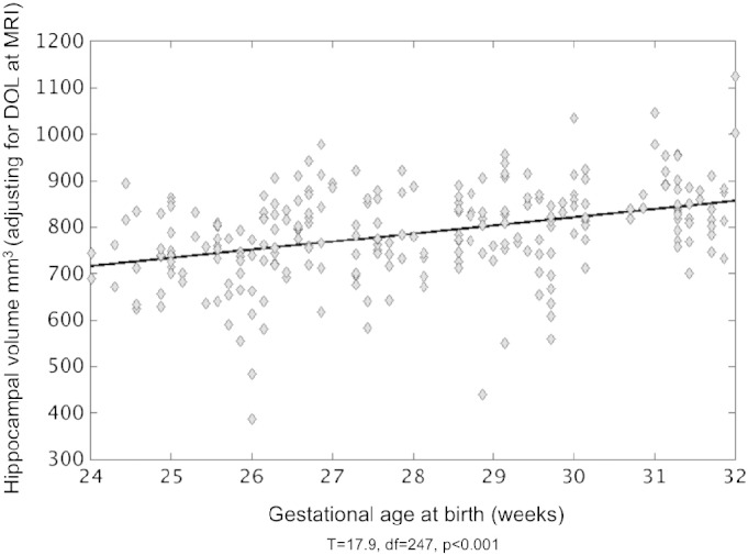
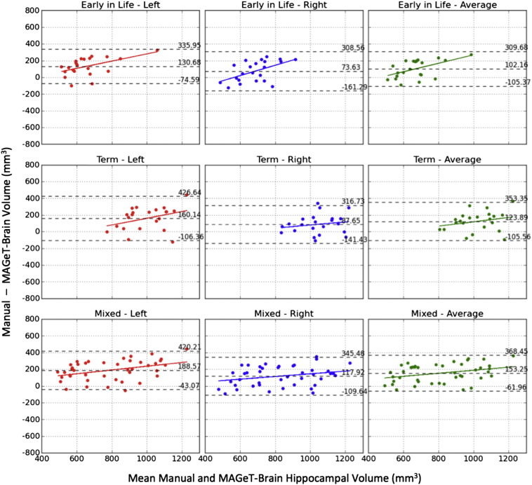
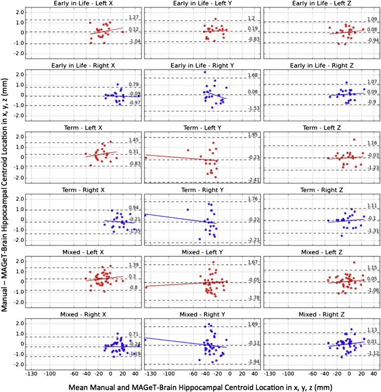
The final segmentations demonstrate that MAGeT-Brain consistently provides accurate segmentations in comparison to manually derived gold standards (mean Dice's Kappa > 0.79 and Euclidean distance <1.3 mm between centroids). Using this method, we demonstrate that the average volume of the hippocampus is significantly different (p < 0.0001) in early-in-life (621.8 mm(3)) and term-equivalent age (958.8 mm(3)). Using these differences, we generalize the hippocampal growth rate to 38.3 ± 11.7 mm(3)/week and 40.5 ± 12.9 mm(3)/week for the left and right hippocampi respectively. Not surprisingly, younger gestational age at birth is associated with smaller volumes of the hippocampi (p = 0.001).
MAGeT-Brain is capable of segmenting hippocampi accurately in preterm neonates, even at early-in-life. Hippocampal asymmetry with a larger right side is demonstrated on early-in-life images, suggesting that this phenomenon has its onset in the 3rd trimester of gestation. Hippocampal volume assessed at the time of early-in-life and term-equivalent age is linearly associated with GA at birth, whereby smaller volumes are associated with earlier birth.
海马体是位于颞叶内侧的一个结构,对学习和记忆至关重要,在早产儿中尤其脆弱。迄今为止,尚未在生命早期(出生后临床状况稳定不久)对早产儿的海马体进行分割。本研究重点关注基于MAGeT-Brain(多个自动生成模板)算法的自动分割方案的开发与验证,该算法用于在早产儿的脑部MRI上勾勒出海马体,这些MRI不仅在足月等效年龄时获取,也在生命早期获取。
首先,我们提出了一个三步手动分割方案来勾勒早产儿的海马体,并将此方案应用于22张生命早期图像和22张足月图像。这些手动分割被视为评估自动分割的金标准。MAGeT-Brain自动海马体分割流程仅需要少量输入图谱,并通过使用中间模板库减少配准和重采样误差。我们在三项验证研究中评估MAGeT-Brain的分割准确性,评估从生命早期到足月等效年龄的海马体生长情况,并研究早产对海马体体积的影响。第一个实验在三组10倍蒙特卡洛交叉验证(MCCV)分析中,使用187组不同的输入图谱和模板,全面验证MAGeT-Brain分割。第二个实验对168张生命早期图像和154张足月图像上的新生儿海马体进行分割,并评估125名婴儿从生命早期到足月等效年龄的海马体生长率。第三个实验使用线性回归分析出生时的孕周(GA)对生命早期和足月等效年龄时海马体平均体积的影响。
最终分割结果表明,与手动得出的金标准相比,MAGeT-Brain始终能提供准确的分割结果(平均骰子系数>0.79,质心之间的欧几里得距离<1.3毫米)。使用此方法,我们证明海马体的平均体积在生命早期(621.8立方毫米)和足月等效年龄(958.8立方毫米)存在显著差异(p<0.0001)。利用这些差异,我们将左右海马体的生长率分别概括为38.3±11.7立方毫米/周和40.5±12.9立方毫米/周。不出所料,出生时孕周越小,海马体体积越小(p = 0.001)。
MAGeT-Brain能够准确分割早产儿的海马体,即使在生命早期也能做到。在生命早期图像上显示出右侧较大的海马体不对称性,这表明这种现象在妊娠晚期开始出现。在生命早期和足月等效年龄时评估的海马体体积与出生时的孕周呈线性相关,孕周越小,出生越早。