Tavakoli Amirrasoul, Hu Shiqiong, Ebrahim Seham, Kachar Bechara
Laboratory of Cell Structure and Dynamics, National Institute on Deafness and Other Communication Disorders, National Institutes of Health, Bethesda, MD 20892, USA.
Center for Membrane and Cell Physiology, Department of Molecular Physiology and Biological Physics, University of Virginia, Charlotteville, VA 22903, USA.
Res Sq. 2024 Oct 21:rs.3.rs-5200876. doi: 10.21203/rs.3.rs-5200876/v1.
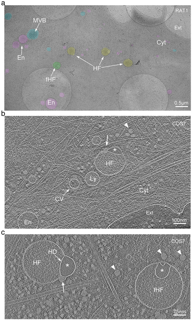
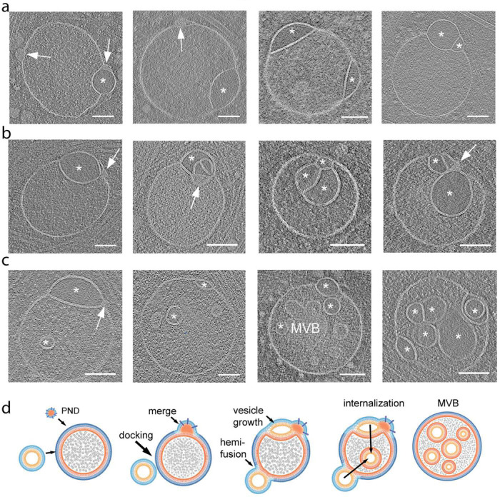
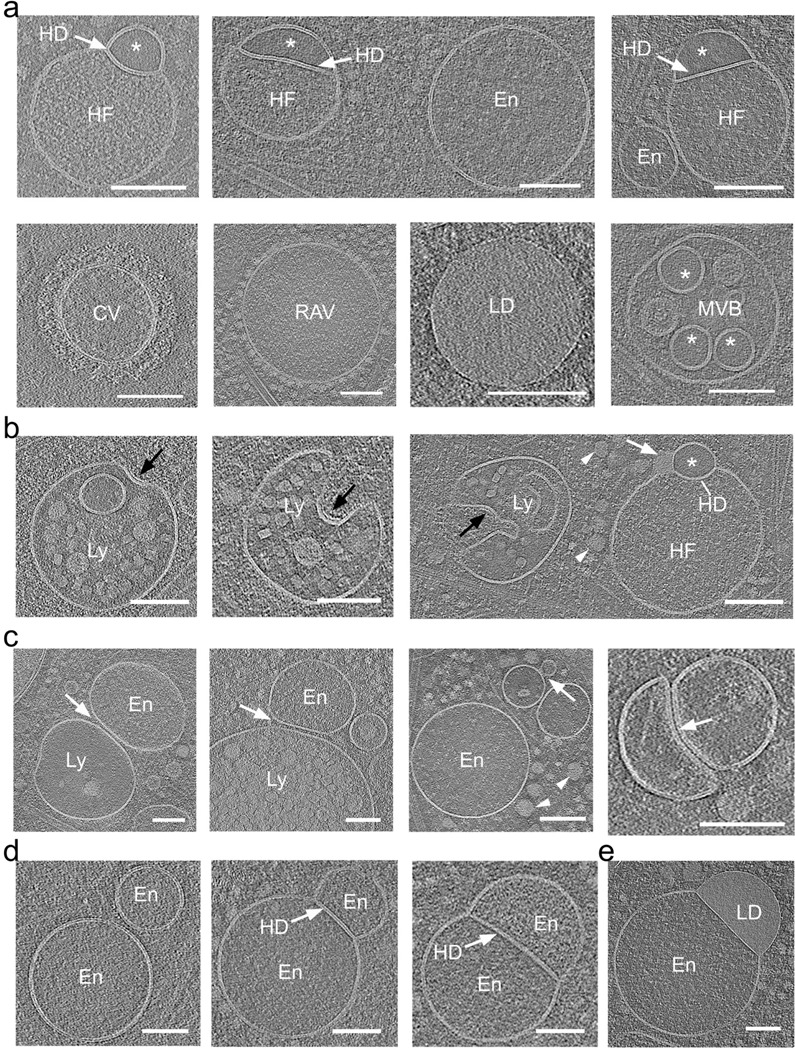
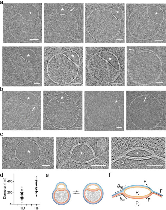
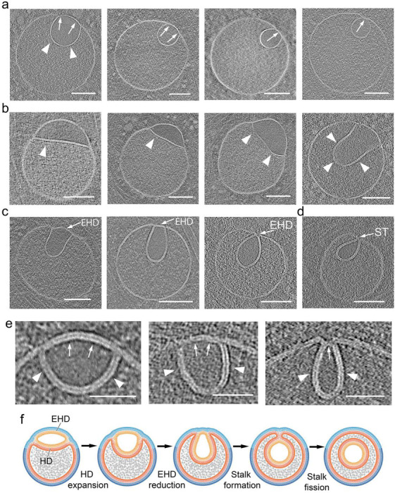
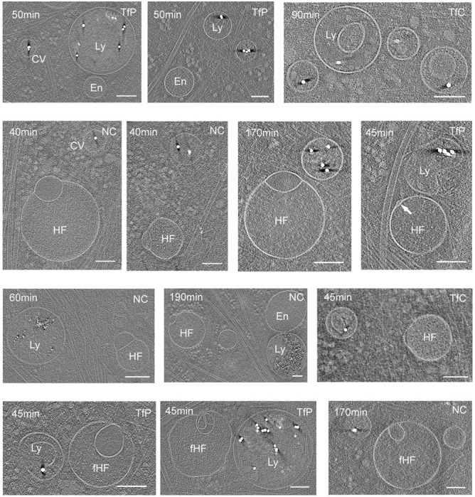
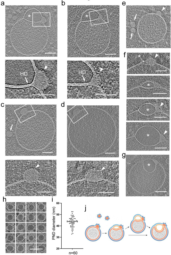
The complex, pleiomorphic membrane structure of the vesicular components within the endolysosomal system has been appreciated through decades of classical electron microscopy. However, due to the heavy fixation and staining required in these approaches, in situ visualization of fragile intermediates between early endosomes, late endosomes and ultimately multivesicular bodies (MVBs), remains elusive, raising the likelihood that other structures may have also been overlooked. Here, using in situ cryo-electron tomography in four mammalian cell lines, we discover heterotypic hemifused vesicles that share an extended hemifusion diaphragm, associated with a 42nm proteolipid nanodroplet (PND). We term this previously undescribed vesicular organelle-complex, "hemifusome". Hemifusomes make up approximately 10% of the organelle pool of the endolysosomal system, but do not participate directly in transferrin-mediated endocytosis. Hemifusomes exist in compound conformations and also contain intraluminal vesicles. Based on their range of morphologies, and the consistent presence of the PND at sites of compound hemifused vesicles, we propose that hemifusomes function as platforms for vesicular biogenesis mediated by the PND. These findings offer direct in situ evidence for a long-lived hemifusion diaphragm, and a new, ESCRT-independent model for the formation of late endosomes containing intraluminal vesicles and ultimately MVBs.
通过数十年的经典电子显微镜观察,人们已经认识到内溶酶体系统中囊泡成分复杂的、多形性的膜结构。然而,由于这些方法需要进行重度固定和染色,早期内体、晚期内体以及最终的多囊泡体(MVB)之间脆弱中间体的原位可视化仍然难以实现,这增加了其他结构可能也被忽视的可能性。在这里,我们使用原位冷冻电子断层扫描技术对四种哺乳动物细胞系进行研究,发现了具有延伸半融合隔膜的异型半融合囊泡,该隔膜与一个42纳米的蛋白脂质纳米液滴(PND)相关联。我们将这种以前未被描述的囊泡细胞器复合体称为“半融合体”。半融合体约占内溶酶体系统细胞器池的10%,但不直接参与转铁蛋白介导的内吞作用。半融合体以复合构象存在,并且还包含腔内囊泡。基于它们的形态范围以及在复合半融合囊泡部位始终存在PND,我们提出半融合体作为由PND介导的囊泡生物发生的平台发挥作用。这些发现为长期存在的半融合隔膜提供了直接的原位证据,并为包含腔内囊泡以及最终MVB的晚期内体形成提供了一种新的、不依赖于ESCRT的模型。