Hu Xinyue, Liu Yali, Tang Bincheng, Hu Jiwei, He Hua, Liu Hehe, Li Liang, Hu Shenqiang, Wang Jiwen
State Key Laboratory of Swine and Poultry Breeding Industry, College of Animal Science and Technology, Sichuan Agricultural University, Chengdu, Sichuan, PR China.
State Key Laboratory of Swine and Poultry Breeding Industry, College of Animal Science and Technology, Sichuan Agricultural University, Chengdu, Sichuan, PR China.
Poult Sci. 2024 Dec;103(12):104498. doi: 10.1016/j.psj.2024.104498. Epub 2024 Nov 2.
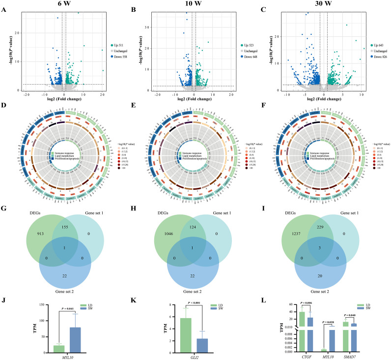
Pectoral muscle development is an important economic trait. According to the different essence, muscle development can be divided into 2 processes: embryonic muscle fiber generation and postnatal muscle fiber hypertrophy, and postnatal muscle fiber hypertrophy has a greater impact on muscle development than the number of muscle fibers formed during the embryonic phase in poultry. However, the underlying mechanisms regulating the hypertrophy of goose pectoral muscles have not been elucidated. Therefore, the purpose of the present study was to conduct transcriptome sequencing in pectoral muscles of both Landes (LD) and Sichuan White (SW) geese at 6, 10, and 30 weeks of age to reveal the molecular mechanisms regulating pectoral muscle hypertrophy through intra-breed and inter-breed bioinformatics analyses. Phenotypically, the pectoral muscle weight/index of LD and SW geese increased from 6 to 30 weeks of age, and except for the pectoral muscle index at 10 weeks of age (P = 0.962), at the same age, the pectoral muscle weight/index of LD geese were significantly higher than that of SW geese (P < 0.05). In transcriptional regulation, intra-breed bioinformatics analysis identified 3331 genes whose expression levels were opposite to the trend of pectoral muscle hypertrophy both in LD and SW geese, and the 3331 genes were mainly enriched into abundant KEGG pathways related to lipid metabolism, proliferation/apoptosis, and immune response. Moreover, 23 genes (including SLC2A10, TNFRSF1A, PRKAA1, SLC27A4, ITGB2, THY1, RHOA, MYL10, ACTB, PRKCB, PIK3R2, RAC2, DMD, LATS2, YAP1, WWTR1, SMAD7, CTGF, FGF1, AXIN2, GLI2, ID2, and CCND2) who were enriched in 6 crosstalk pathways named viral myocarditis, insulin resistance, sphingolipid signaling pathway, hippo signaling pathway, chemokine signaling pathway, and leukocyte transendothelial migration were identified as the key candidate genes regulating the hypertrophy of goose pectoral muscles. In inter-breed bioinformatics analysis, abundant different expression genes (DEGs) related to lipid metabolism, immune response, and proliferation/apoptosis were identified between LD and SW geese too, and compared with SW geese, the expression level of MYL10 in LD geese was lower, while the expression levels of GLI2/CTGF/SMAD7 in LD geese were higher. These results suggested that the hypertrophy of goose pectoral muscles might be achieved through more lipid deposition and less leukocyte infiltration to promote the proliferation of cells within the muscles, and the low expression of MYL10 and high expressions of GLI2/CTGF/SMAD7 might the keys to induce the pectoral muscle hypertrophy of LD geese from 6 to 30 weeks of age over that of SW geese. All data the present study obtained will provide new insights into the molecular mechanisms regulating the hypertrophy of goose pectoral muscles.
胸肌发育是一个重要的经济性状。根据不同本质,肌肉发育可分为两个过程:胚胎期肌纤维生成和出生后肌纤维肥大,在家禽中,出生后肌纤维肥大对肌肉发育的影响比胚胎期形成的肌纤维数量更大。然而,调控鹅胸肌肥大的潜在机制尚未阐明。因此,本研究的目的是对朗德鹅(LD)和四川白鹅(SW)6周龄、10周龄和30周龄时的胸肌进行转录组测序,通过品种内和品种间的生物信息学分析揭示调控胸肌肥大的分子机制。表型上,LD和SW鹅的胸肌重量/指数从6周龄到30周龄增加,除10周龄时的胸肌指数外(P = 0.962),在相同年龄,LD鹅的胸肌重量/指数显著高于SW鹅(P < 0.05)。在转录调控方面,品种内生物信息学分析在LD和SW鹅中均鉴定出3331个基因,其表达水平与胸肌肥大趋势相反,这3331个基因主要富集到与脂质代谢、增殖/凋亡和免疫反应相关的大量KEGG通路中。此外,在名为病毒性心肌炎、胰岛素抵抗、鞘脂信号通路、河马信号通路、趋化因子信号通路和白细胞跨内皮迁移的6条相互作用通路中富集的23个基因(包括SLC2A10、TNFRSF1A、PRKAA1、SLC27A4、ITGB2、THY1、RHOA、MYL10、ACTB、PRKCB、PIK3R2、RAC2、DMD、LATS2、YAP1、WWTR1、SMAD7、CTGF、FGF1、AXIN2、GLI2、ID2和CCND2)被确定为调控鹅胸肌肥大的关键候选基因。在品种间生物信息学分析中,LD和SW鹅之间也鉴定出大量与脂质代谢、免疫反应和增殖/凋亡相关的差异表达基因(DEG),与SW鹅相比,LD鹅中MYL10的表达水平较低,而GLI2/CTGF/SMAD7在LD鹅中的表达水平较高。这些结果表明,鹅胸肌肥大可能是通过更多的脂质沉积和更少的白细胞浸润来促进肌肉内细胞增殖实现的,MYL10的低表达和GLI2/CTGF/SMAD7的高表达可能是6至30周龄LD鹅胸肌肥大超过SW鹅的关键。本研究获得的所有数据将为调控鹅胸肌肥大的分子机制提供新的见解。