Department of Radiation Oncology, National Cancer Center/National Clinical Research Center for Cancer/Cancer Hospital, Chinese Academy of Medical Sciences and Peking Union Medical College, Beijing, 100021, China.
Department of Radiation Oncology, Clinical Oncology School of Fujian Medical University, Fujian Cancer Hospital, Fuzhou, 350014, China.
BMC Med Imaging. 2024 Nov 18;24(1):312. doi: 10.1186/s12880-024-01469-0.
Tumor bed (TB) is the residual cavity of resected tumor after surgery. Delineating TB from CT is crucial in generating clinical target volume for radiotherapy. Due to multiple surgical effects and low image contrast, segmenting TB from soft tissue is challenging. In clinical practice, titanium clips were used as marks to guide the searching of TB. However, this information is limited and may cause large error. To provide more prior location information, the tumor regions on both pre-operative and post-operative CTs are both used by the deep learning model in segmenting TB from surrounding tissues.
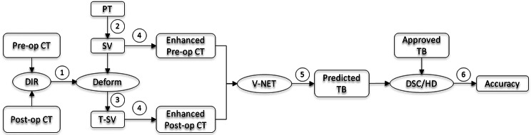
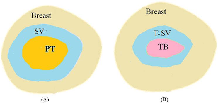
For breast cancer patient after surgery and going to be treated by radiotherapy, it is important to delineate the target volume for treatment planning. In clinical practice, the target volume is usually generated from TB by adding a certain margin. Therefore, it is crucial to identify TB from soft tissue. To facilitate this process, a deep learning model is developed to segment TB from CT with the guidance of prior tumor location. Initially, the tumor contour on the pre-operative CT is delineated by physician for surgical planning purpose. Then this contour is transformed to the post-operative CT via the deformable image registration between paired pre-operative and post-operative CTs. The original and transformed tumor regions are both used as inputs for predicting the possible region of TB by the deep-learning model.
Compared to the one without prior tumor contour information, the dice similarity coefficient of the deep-learning model with the prior tumor contour information is improved significantly (0.812 vs. 0.520, P = 0.001). Compared to the traditional gray-level thresholding method, the dice similarity coefficient of the deep-learning model with the prior tumor contour information is improved significantly (0.812 vs.0.633, P = 0.0005).
The prior tumor contours on both pre-operative and post-operative CTs provide valuable information in searching for the precise location of TB on post-operative CT. The proposed method provided a feasible way to assist auto-segmentation of TB in treatment planning of radiotherapy after breast-conserving surgery.
肿瘤床(TB)是手术后切除肿瘤的残余腔。从 CT 中勾画 TB 对于生成放射治疗的临床靶区至关重要。由于多种手术影响和低图像对比度,从软组织中分割 TB 具有挑战性。在临床实践中,钛夹被用作标记物来指导 TB 的搜索。然而,这种信息是有限的,可能会导致较大的误差。为了提供更多的先验位置信息,深度学习模型同时使用术前和术后 CT 上的肿瘤区域来从周围组织中分割 TB。
对于手术后准备接受放射治疗的乳腺癌患者,勾画靶区进行治疗计划非常重要。在临床实践中,靶区通常是通过在 TB 上添加一定的边界来生成的。因此,从软组织中识别 TB 至关重要。为了方便这一过程,开发了一种深度学习模型,通过先验肿瘤位置的指导来从 CT 中分割 TB。首先,医生为手术计划目的在术前 CT 上勾画肿瘤轮廓。然后,通过配准术前和术后 CT 之间的变形图像,将该轮廓转换到术后 CT 上。原始和转换后的肿瘤区域都作为输入,由深度学习模型预测 TB 的可能区域。
与没有先验肿瘤轮廓信息的模型相比,具有先验肿瘤轮廓信息的深度学习模型的 Dice 相似系数显著提高(0.812 比 0.520,P=0.001)。与传统的灰度阈值法相比,具有先验肿瘤轮廓信息的深度学习模型的 Dice 相似系数显著提高(0.812 比 0.633,P=0.0005)。
术前和术后 CT 上的先验肿瘤轮廓为在术后 CT 上搜索 TB 的精确位置提供了有价值的信息。该方法为保乳手术后放射治疗的 TB 自动分割提供了一种可行的方法。