• 文献检索
  • 文档翻译
  • 深度研究
  • 学术资讯
  • Suppr Zotero 插件Zotero 插件
  • 邀请有礼
  • 套餐&价格
  • 历史记录
应用&插件
Suppr Zotero 插件Zotero 插件浏览器插件Mac 客户端Windows 客户端微信小程序
定价
高级版会员购买积分包购买API积分包
服务
文献检索文档翻译深度研究API 文档MCP 服务
关于我们
关于 Suppr公司介绍联系我们用户协议隐私条款
关注我们

Suppr 超能文献

核心技术专利:CN118964589B侵权必究
粤ICP备2023148730 号-1Suppr @ 2026

文献检索

告别复杂PubMed语法,用中文像聊天一样搜索,搜遍4000万医学文献。AI智能推荐,让科研检索更轻松。

立即免费搜索

文件翻译

保留排版,准确专业,支持PDF/Word/PPT等文件格式,支持 12+语言互译。

免费翻译文档

深度研究

AI帮你快速写综述,25分钟生成高质量综述,智能提取关键信息,辅助科研写作。

立即免费体验

人玻璃体细胞的短期培养保留了其初始表型,并显示出其收缩能力。

Short-Term Culture of Human Hyalocytes Retains Their Initial Phenotype and Displays Their Contraction Abilities.

机构信息

Research and Development Laboratory for Biochemical, Molecular and Cellular Applications in Ophthalmological Science, IRCCS-Fondazione Bietti, 00184 Rome, Italy.

Surgical Retina Research Unit, IRCCS-Fondazione Bietti, 00184 Rome, Italy.

出版信息

Cells. 2024 Nov 6;13(22):1837. doi: 10.3390/cells13221837.

DOI:10.3390/cells13221837
PMID:39594586
原文链接:https://pmc.ncbi.nlm.nih.gov/articles/PMC11592754/
Abstract

BACKGROUND

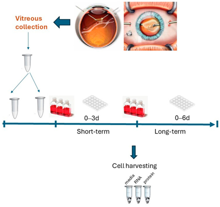
Hyalocytes are the main vitreal cell types with critical functions in health and vitreoretinal diseases. Our aim was to develop cultures of human hyalocytes and verify the retention of their initial cellular features after 3 and 6 days of culturing (3 d and 6 d) by analyzing and comparing a few morphological and functional parameters.

METHODS

Vitreous samples (n = 22) were collected and vitreous cells and bead-enriched hyalocytes were developed and compared (3 d vs. 6 d cultures). Vitreous and conditioned media were tested for collagen, vascular endothelial growth factor (VEGF), transforming growth factor β1 (TGFβ1), nerve growth factor (NGF), matrix metalloproteinases (MMPs)/tissue inhibitors of metalloproteinases (TIMPs) and alpha-smooth muscle actin (αSMA) expression (ELISA, array/IP/WB, RT-PCR). Cells were observed at light and fluorescent microscopy (phenotypical properties) and tested for their 3D collagen gel contraction abilities.

RESULTS

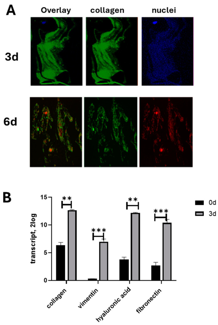
An increased expression of collagens, vimentin, fibronectin, and the MMP9/TIMP1 ratio were observed in vitreous tissues. In 3 d cultures, collagens and MMP9 were upregulated while the related tissue-enzymes were deregulated. Vitreous samples also showed high levels of TGFβ1, VEGF, and NGF, and this protein signature was retained at 3 d while decreased at 6 d. The original phenotype (low αSMA) was retained at 3 d from seeding while an increased αSMA expression was observed at 6 d; NGF/trkA was expressed in cultured hyalocytes and partially drives the collagen retraction.

CONCLUSIONS

The vitreous print comparison between untouched and cultured hyalocytes allowed us, on one side, to select 3 d cultures and, on the other, to highlight the neuroprotective/contractile NGF in vitro hyalocytes effects. The possibility of scoring reactive hyalocytes would represent an interesting aspect of screening the vitreoretinal interface severity.

摘要

背景

玻璃体细胞是主要的玻璃体细胞类型,在健康和玻璃体视网膜疾病中具有关键功能。我们的目的是培养人眼玻璃体细胞,并通过分析和比较一些形态和功能参数,验证在培养 3 天和 6 天后(3d 和 6d)它们是否保留了最初的细胞特征。

方法

收集玻璃体样本(n=22),并开发玻璃体细胞和珠富集的玻璃体细胞,并进行比较(3d 与 6d 培养)。测试玻璃体和条件培养基中胶原蛋白、血管内皮生长因子(VEGF)、转化生长因子β1(TGFβ1)、神经生长因子(NGF)、基质金属蛋白酶(MMPs)/金属蛋白酶组织抑制剂(TIMPs)和α-平滑肌肌动蛋白(αSMA)的表达(ELISA、阵列/免疫沉淀/WB、RT-PCR)。通过荧光显微镜观察细胞(表型特性),并测试其 3D 胶原凝胶收缩能力。

结果

在玻璃体组织中观察到胶原蛋白、波形蛋白、纤维连接蛋白和 MMP9/TIMP1 比值的表达增加。在 3d 培养物中,胶原蛋白和 MMP9 上调,而相关的组织酶则失调。玻璃体样本还显示出高水平的 TGFβ1、VEGF 和 NGF,这种蛋白质特征在 3d 时保留,而在 6d 时降低。在接种后 3d,原始表型(低αSMA)得以保留,而在 6d 时观察到αSMA 表达增加;培养的玻璃体细胞中表达了 NGF/trkA,并部分驱动胶原收缩。

结论

未经处理和培养的玻璃体细胞之间的玻璃体印迹比较,使我们一方面能够选择 3d 培养物,另一方面突出体外玻璃体细胞的神经保护/收缩作用的 NGF。对反应性玻璃体细胞进行评分的可能性将是评估玻璃体视网膜界面严重程度的一个有趣方面。

https://cdn.ncbi.nlm.nih.gov/pmc/blobs/fb0d/11592754/e1b811f1d30a/cells-13-01837-g007.jpg
https://cdn.ncbi.nlm.nih.gov/pmc/blobs/fb0d/11592754/1db73b23e76d/cells-13-01837-g001.jpg
https://cdn.ncbi.nlm.nih.gov/pmc/blobs/fb0d/11592754/b1b7a8b8264a/cells-13-01837-g002.jpg
https://cdn.ncbi.nlm.nih.gov/pmc/blobs/fb0d/11592754/571195640b0c/cells-13-01837-g003.jpg
https://cdn.ncbi.nlm.nih.gov/pmc/blobs/fb0d/11592754/e19cd36c4dd5/cells-13-01837-g004.jpg
https://cdn.ncbi.nlm.nih.gov/pmc/blobs/fb0d/11592754/66eb8479393a/cells-13-01837-g005.jpg
https://cdn.ncbi.nlm.nih.gov/pmc/blobs/fb0d/11592754/2fc74675bc44/cells-13-01837-g006.jpg
https://cdn.ncbi.nlm.nih.gov/pmc/blobs/fb0d/11592754/e1b811f1d30a/cells-13-01837-g007.jpg

相似文献

1
Short-Term Culture of Human Hyalocytes Retains Their Initial Phenotype and Displays Their Contraction Abilities.人玻璃体细胞的短期培养保留了其初始表型,并显示出其收缩能力。
Cells. 2024 Nov 6;13(22):1837. doi: 10.3390/cells13221837.
2
The involvement of the rho-kinase pathway and its regulation in cytokine-induced collagen gel contraction by hyalocytes.Rho激酶通路的参与及其在透明细胞介导的细胞因子诱导的胶原凝胶收缩中的调节作用。
Invest Ophthalmol Vis Sci. 2004 Nov;45(11):3896-903. doi: 10.1167/iovs.03-1330.
3
Possible contribution of hyalocytes to idiopathic epiretinal membrane formation and its contraction.玻璃体细胞对特发性视网膜前膜形成及其收缩的可能作用。
Br J Ophthalmol. 2009 Aug;93(8):1020-6. doi: 10.1136/bjo.2008.155069. Epub 2009 May 7.
4
Nerve growth factor has a modulatory role on human primary fibroblast cultures derived from vernal keratoconjunctivitis-affected conjunctiva.神经生长因子对源自春季角结膜炎累及结膜的人原代成纤维细胞培养物具有调节作用。
Mol Vis. 2007 Jun 21;13:981-7.
5
[The effect of OPTC-shRNA on the bovine retinal pigment epithelial cells and hyalocytes co-culture collagen gel contraction system].[OPTC-shRNA对牛视网膜色素上皮细胞与玻璃体细胞共培养胶原凝胶收缩系统的影响]
Zhonghua Yan Ke Za Zhi. 2013 Feb;49(2):155-62.
6
In vitro generation of primary cultures of human hyalocytes.人玻璃体细胞原代培养物的体外生成。
Mol Vis. 2020 Dec 30;26:818-829. eCollection 2020.
7
Hyalocytes-guardians of the vitreoretinal interface.玻璃体细胞——视网膜界面的守护者。
Graefes Arch Clin Exp Ophthalmol. 2024 Sep;262(9):2765-2784. doi: 10.1007/s00417-024-06448-3. Epub 2024 Apr 3.
8
Hyalocytes in idiopathic epiretinal membranes: a correlative light and electron microscopic study.特发性视网膜前膜中的玻璃体细胞:光镜与电镜相关研究
Graefes Arch Clin Exp Ophthalmol. 2014 Dec;252(12):1887-94. doi: 10.1007/s00417-014-2841-x. Epub 2014 Nov 7.
9
The in vitro response of human retinal endothelial cells to cytokines and other chemically active agents is altered by coculture with vitreous-derived hyalocytes.人视网膜血管内皮细胞对细胞因子和其他化学活性物质的体外反应,会受到与玻璃体液来源的玻璃体细胞共培养的影响而发生改变。
Acta Ophthalmol. 2010 May;88(3):e66-72. doi: 10.1111/j.1755-3768.2010.01879.x. Epub 2010 Mar 23.
10
Functional properties of hyalocytes under PDGF-rich conditions.富含血小板源性生长因子(PDGF)条件下玻璃体细胞的功能特性。
Invest Ophthalmol Vis Sci. 2004 Jul;45(7):2107-14. doi: 10.1167/iovs.03-1092.

引用本文的文献

1
Expression of Reelin, Aβ1-42, Tau and FTH1 in Idiopathic Epiretinal Membranes: Exploring the Link Between Reelin and Neurodegenerative Biomarkers.视网膜前膜中Reelin、Aβ1-42、Tau和FTH1的表达:探索Reelin与神经退行性生物标志物之间的联系
Biomolecules. 2025 Aug 18;15(8):1187. doi: 10.3390/biom15081187.
2
Differentiation of human hyalocytes from induced pluripotent stem cells through ascorbic acid treatment.通过抗坏血酸处理从诱导多能干细胞分化出人类玻璃体细胞。
Hum Cell. 2025 Feb 12;38(2):52. doi: 10.1007/s13577-025-01182-2.

本文引用的文献

1
TLR2 and TLR4 Are Expressed in Epiretinal Membranes: Possible Links with Vitreous Levels of Complement Fragments and DAMP-Related Proteins.TLR2 和 TLR4 在视网膜前膜中表达:与玻璃体液中补体片段和 DAMPs 相关蛋白的可能联系。
Int J Mol Sci. 2024 Jul 15;25(14):7732. doi: 10.3390/ijms25147732.
2
Redefining the ontogeny of hyalocytes as yolk sac-derived tissue-resident macrophages of the vitreous body.重新定义玻璃体内的原纤维细胞为卵黄囊来源的组织驻留巨噬细胞。
J Neuroinflammation. 2024 Jul 3;21(1):168. doi: 10.1186/s12974-024-03110-x.
3
Hyalocytes-guardians of the vitreoretinal interface.
玻璃体细胞——视网膜界面的守护者。
Graefes Arch Clin Exp Ophthalmol. 2024 Sep;262(9):2765-2784. doi: 10.1007/s00417-024-06448-3. Epub 2024 Apr 3.
4
Effect of Fetal Bovine Serum or Basic Fibroblast Growth Factor on Cell Survival and the Proliferation of Neural Stem Cells: The Influence of Homocysteine Treatment.胎牛血清或碱性成纤维细胞生长因子对神经干细胞存活及增殖的影响:同型半胱氨酸处理的作用
Int J Mol Sci. 2023 Sep 15;24(18):14161. doi: 10.3390/ijms241814161.
5
Role of the Vitreous in Retinal Pathology: A Narrative Review.玻璃体在视网膜病理学中的作用:一篇叙述性综述。
Cureus. 2023 Aug 23;15(8):e43990. doi: 10.7759/cureus.43990. eCollection 2023 Aug.
6
Immune Fingerprint in Diabetes: Ocular Surface and Retinal Inflammation.糖尿病的免疫指纹:眼表和视网膜炎症。
Int J Mol Sci. 2023 Jun 6;24(12):9821. doi: 10.3390/ijms24129821.
7
Experimental Models to Study Epithelial-Mesenchymal Transition in Proliferative Vitreoretinopathy.研究增生性玻璃体视网膜病变中上皮-间充质转化的实验模型。
Int J Mol Sci. 2023 Feb 24;24(5):4509. doi: 10.3390/ijms24054509.
8
A Beginner's Guide to Cell Culture: Practical Advice for Preventing Needless Problems.细胞培养新手指南:预防不必要问题的实用建议。
Cells. 2023 Feb 21;12(5):682. doi: 10.3390/cells12050682.
9
Vitreous Substitutes from Bench to the Operating Room in a Translational Approach: Review and Future Endeavors in Vitreoretinal Surgery.从基础到手术:转化研究视角下的玻璃体替代物在眼后段手术中的应用:综述与未来展望。
Int J Mol Sci. 2023 Feb 7;24(4):3342. doi: 10.3390/ijms24043342.
10
Hyalocyte origin, structure, and imaging.玻璃体细胞的起源、结构与成像。
Expert Rev Ophthalmol. 2022;17(4):233-248. doi: 10.1080/17469899.2022.2100762. Epub 2022 Sep 6.