Qian Meizhen, Wang Jianbao, Gao Yang, Chen Ming, Liu Yin, Zhou Dengfeng, Lu Haidong D, Zhang Xiaotong, Hu Jia Ming, Roe Anna Wang
Department of Neurosurgery of the Second Affiliated Hospital & Liangzhu Laboratory of Zhejiang University School of Medicine, Zhejiang University, Hangzhou, China.
Interdisciplinary Institute of Neuroscience and Technology, School of Medicine, Zhejiang University, Hangzhou, China.
Nat Neurosci. 2025 Jan;28(1):137-149. doi: 10.1038/s41593-024-01810-4. Epub 2024 Dec 5.
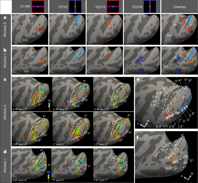
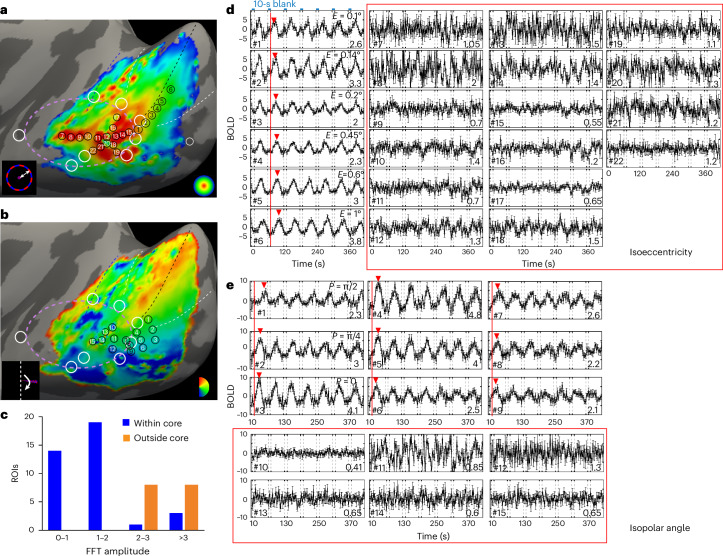
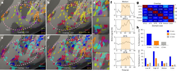
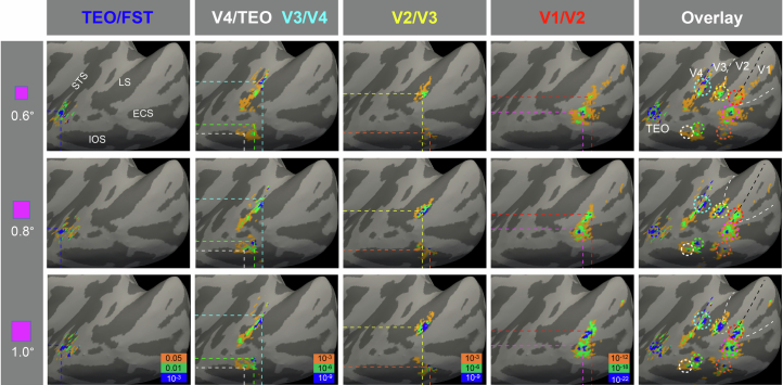
In humans and nonhuman primates, the central 1° of vision is processed by the foveola, a retinal structure that comprises a high density of photoreceptors and is crucial for primate-specific high-acuity vision, color vision and gaze-directed visual attention. Here, we developed high-spatial-resolution ultrahigh-field 7T functional magnetic resonance imaging methods for functional mapping of the foveolar visual cortex in awake monkeys. In the ventral pathway (visual areas V1-V4 and the posterior inferior temporal cortex), viewing of a small foveolar spot elicits a ring of multiple (eight) foveolar representations per hemisphere. This ring surrounds an area called the 'foveolar core', which is populated by millimeter-scale functional domains sensitive to fine stimuli and high spatial frequencies, consistent with foveolar visual acuity, color and achromatic information and motion. Thus, this elaborate rerepresentation of central vision coupled with a previously unknown foveolar core area signifies a cortical specialization for primate foveation behaviors.
在人类和非人类灵长类动物中,中央1°视野由中央凹处理,中央凹是一种视网膜结构,包含高密度的光感受器,对于灵长类动物特有的高敏锐度视觉、色觉和凝视导向视觉注意力至关重要。在此,我们开发了高空间分辨率超高场7T功能磁共振成像方法,用于在清醒猴子中对中央凹视觉皮层进行功能映射。在腹侧通路(视觉区域V1-V4和颞下后皮质)中,观察一个小的中央凹光斑会在每个半球引发多个(八个)中央凹表征的环。这个环围绕着一个称为“中央凹核心”的区域,该区域由对精细刺激和高空间频率敏感的毫米级功能域组成,与中央凹视觉敏锐度、颜色和非彩色信息以及运动一致。因此,这种对中央视觉的精细重新表征以及一个先前未知的中央凹核心区域表明了灵长类动物中央凹行为的皮质特化。