• 文献检索
  • 文档翻译
  • 深度研究
  • 学术资讯
  • Suppr Zotero 插件Zotero 插件
  • 邀请有礼
  • 套餐&价格
  • 历史记录
应用&插件
Suppr Zotero 插件Zotero 插件浏览器插件Mac 客户端Windows 客户端微信小程序
定价
高级版会员购买积分包购买API积分包
服务
文献检索文档翻译深度研究API 文档MCP 服务
关于我们
关于 Suppr公司介绍联系我们用户协议隐私条款
关注我们

Suppr 超能文献

核心技术专利:CN118964589B侵权必究
粤ICP备2023148730 号-1Suppr @ 2026

文献检索

告别复杂PubMed语法,用中文像聊天一样搜索,搜遍4000万医学文献。AI智能推荐,让科研检索更轻松。

立即免费搜索

文件翻译

保留排版,准确专业,支持PDF/Word/PPT等文件格式,支持 12+语言互译。

免费翻译文档

深度研究

AI帮你快速写综述,25分钟生成高质量综述,智能提取关键信息,辅助科研写作。

立即免费体验

通过可解释深度学习利用光学相干断层扫描成像自动检测视网膜中央动脉阻塞

Automated Detection of Central Retinal Artery Occlusion Using OCT Imaging via Explainable Deep Learning.

作者信息

Beuse Ansgar, Wenzel Daniel Alexander, Spitzer Martin Stephan, Bartz-Schmidt Karl Ulrich, Schultheiss Maximilian, Poli Sven, Grohmann Carsten

机构信息

Department of Ophthalmology, University Medical Center Hamburg-Eppendorf, Hamburg, Germany.

University Eye Hospital, Centre for Ophthalmology, University Hospital Tübingen, Tübingen, Germany.

出版信息

Ophthalmol Sci. 2024 Oct 16;5(2):100630. doi: 10.1016/j.xops.2024.100630. eCollection 2025 Mar-Apr.

DOI:10.1016/j.xops.2024.100630
PMID:39669299
原文链接:https://pmc.ncbi.nlm.nih.gov/articles/PMC11634984/
Abstract

OBJECTIVE

To demonstrate the capability of a deep learning model to detect central retinal artery occlusion (CRAO), a retinal pathology with significant clinical urgency, using OCT data.

DESIGN

Retrospective, external validation study analyzing OCT and clinical baseline data of 2 institutions via deep learning classification analysis.

SUBJECTS

Patients presenting to the University Medical Center Tübingen and the University Medical Center Hamburg-Eppendorf in Germany.

METHODS

OCT data of patients suffering from CRAO, differential diagnosis with (sub) acute visual loss (central retinal vein occlusion, diabetic macular edema, nonarteritic ischemic optic neuropathy), and from controls were expertly graded and distinguished into 3 groups. Our methodological approach involved a nested multiclass five fold cross-validation classification scheme.

MAIN OUTCOME MEASURES

Area under the curve (AUC).

RESULTS

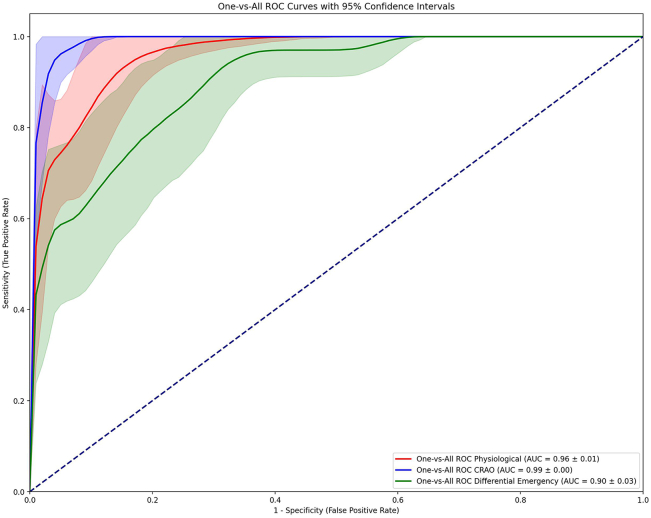
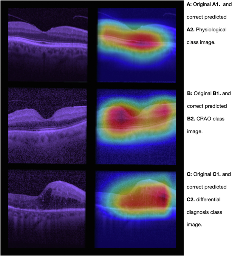
The optimal performance of our algorithm was observed using 30 epochs, complemented by an early stopping mechanism to prevent overfitting. Our model followed a multiclass approach, distinguishing among the 3 different classes: control, CRAO, and differential diagnoses. The evaluation was conducted by the "one vs. all" area under the receiver operating characteristics curve (AUC) method. The results demonstrated AUC of 0.96 (95% confidence interval [CI], ± 0.01); 0.99 (95% CI, ± 0.00); and 0.90 (95% CI, ± 0.03) for each class, respectively.

CONCLUSIONS

Our machine learning algorithm (MLA) exhibited a high AUC, as well as sensitivity and specificity in detecting CRAO and the differential classes, respectively. These findings underscore the potential for deploying MLAs in the identification of less common etiologies within an acute emergency clinical setting.

FINANCIAL DISCLOSURES

Proprietary or commercial disclosure may be found in the Footnotes and Disclosures at the end of this article.

摘要

目的

利用光学相干断层扫描(OCT)数据,证明深度学习模型检测视网膜中央动脉阻塞(CRAO)的能力,CRAO是一种具有重大临床紧迫性的视网膜病变。

设计

通过深度学习分类分析对两个机构的OCT和临床基线数据进行回顾性外部验证研究。

研究对象

德国图宾根大学医学中心和汉堡-埃彭多夫大学医学中心的患者。

方法

对患有CRAO、伴有(亚)急性视力丧失的鉴别诊断(视网膜中央静脉阻塞、糖尿病性黄斑水肿、非动脉性缺血性视神经病变)的患者以及对照组的OCT数据进行专业分级,并分为3组。我们的方法采用了嵌套多类五折交叉验证分类方案。

主要观察指标

曲线下面积(AUC)。

结果

使用30个轮次观察到我们算法的最佳性能,并辅以早期停止机制以防止过拟合。我们的模型采用多类方法,区分3种不同类别:对照组、CRAO组和鉴别诊断组。通过“一对所有”的受试者操作特征曲线(AUC)下面积方法进行评估。结果显示,每组的AUC分别为0.96(95%置信区间[CI],±0.01);0.99(95%CI,±0.00);和0.90(95%CI,±0.03)。

结论

我们的机器学习算法(MLA)在检测CRAO及其鉴别类别时分别表现出高AUC以及敏感性和特异性。这些发现强调了在急性紧急临床环境中部署MLA以识别罕见病因的潜力。

财务披露

专有或商业披露信息可在本文末尾的脚注和披露部分中找到。

https://cdn.ncbi.nlm.nih.gov/pmc/blobs/80db/11634984/4834df9b11ea/gr2.jpg
https://cdn.ncbi.nlm.nih.gov/pmc/blobs/80db/11634984/d90c7970ce16/gr1.jpg
https://cdn.ncbi.nlm.nih.gov/pmc/blobs/80db/11634984/4834df9b11ea/gr2.jpg
https://cdn.ncbi.nlm.nih.gov/pmc/blobs/80db/11634984/d90c7970ce16/gr1.jpg
https://cdn.ncbi.nlm.nih.gov/pmc/blobs/80db/11634984/4834df9b11ea/gr2.jpg

相似文献

1
Automated Detection of Central Retinal Artery Occlusion Using OCT Imaging via Explainable Deep Learning.通过可解释深度学习利用光学相干断层扫描成像自动检测视网膜中央动脉阻塞
Ophthalmol Sci. 2024 Oct 16;5(2):100630. doi: 10.1016/j.xops.2024.100630. eCollection 2025 Mar-Apr.
2
Inner Retinal Layer Hyperreflectivity Is an Early Biomarker for Acute Central Retinal Artery Occlusion.视网膜内层高反射性是急性视网膜中央动脉阻塞的早期生物标志物。
Front Med (Lausanne). 2022 Jul 6;9:854288. doi: 10.3389/fmed.2022.854288. eCollection 2022.
3
Fully Automated Detection and Quantification of Macular Fluid in OCT Using Deep Learning.基于深度学习的 OCT 中黄斑区液全自动化检测和定量分析
Ophthalmology. 2018 Apr;125(4):549-558. doi: 10.1016/j.ophtha.2017.10.031. Epub 2017 Dec 8.
4
Deep Learning Models for the Screening of Cognitive Impairment Using Multimodal Fundus Images.深度学习模型在多模态眼底图像认知障碍筛查中的应用。
Ophthalmol Retina. 2024 Jul;8(7):666-677. doi: 10.1016/j.oret.2024.01.019. Epub 2024 Jan 26.
5
Retinal and choroidal changes and visual outcome in central retinal artery occlusion: an optical coherence tomography study.视网膜中央动脉阻塞时的视网膜和脉络膜变化及视觉预后:一项光学相干断层扫描研究
Am J Ophthalmol. 2015 Apr;159(4):667-76. doi: 10.1016/j.ajo.2015.01.001. Epub 2015 Jan 9.
6
Detection of Macular Neovascularization in Eyes Presenting with Macular Edema using OCT Angiography and a Deep Learning Model.使用光学相干断层扫描血管造影和深度学习模型检测出现黄斑水肿的眼睛中的黄斑新生血管形成
Ophthalmol Retina. 2025 Apr;9(4):378-385. doi: 10.1016/j.oret.2024.10.017. Epub 2024 Oct 24.
7
Ocular vascular occlusive disorders: natural history of visual outcome.眼部血管阻塞性疾病:视力预后的自然史。
Prog Retin Eye Res. 2014 Jul;41:1-25. doi: 10.1016/j.preteyeres.2014.04.001. Epub 2014 Apr 21.
8
Clinical application of multicolor scanning laser ophthalmology in diagnosis and grading of central retinal artery occlusion.多色扫描激光眼科技术在视网膜中央动脉阻塞诊断及分级中的临床应用
Front Neurosci. 2024 Apr 10;18:1327806. doi: 10.3389/fnins.2024.1327806. eCollection 2024.
9
Fully automated detection of retinal disorders by image-based deep learning.基于图像的深度学习技术对视网膜疾病进行全自动检测。
Graefes Arch Clin Exp Ophthalmol. 2019 Mar;257(3):495-505. doi: 10.1007/s00417-018-04224-8. Epub 2019 Jan 4.
10
Deep-Reticular Pseudodrusen-Net: A 3-Dimensional Deep Network for Detection of Reticular Pseudodrusen on OCT Scans.深度网状假性玻璃膜疣网络:一种用于在光学相干断层扫描(OCT)图像上检测网状假性玻璃膜疣的三维深度网络。
Ophthalmol Sci. 2024 Nov 19;5(2):100655. doi: 10.1016/j.xops.2024.100655. eCollection 2025 Mar-Apr.

引用本文的文献

1
Deep learning detection of retinal detachment: Optical coherence tomography staging and estimation of duration of macular detachment.深度学习检测视网膜脱离:光学相干断层扫描分期及黄斑脱离持续时间估计
PLoS One. 2025 Sep 2;20(9):e0329951. doi: 10.1371/journal.pone.0329951. eCollection 2025.
2
Atrial fibrillation in Retinal Artery Occlusions.视网膜动脉阻塞中的心房颤动
Cardiol Cardiovasc Med. 2025;9(4):234-247. Epub 2025 Jul 7.

本文引用的文献

1
Early REperfusion therapy with intravenous alteplase for recovery of VISION in acute central retinal artery occlusion (REVISION): Study protocol of a phase III trial.急性中央视网膜动脉阻塞患者的早期静脉内阿替普酶再灌注治疗以恢复视力(REVISION):一项 III 期试验的研究方案。
Int J Stroke. 2024 Aug;19(7):823-829. doi: 10.1177/17474930241248516. Epub 2024 Jun 25.
2
Application of a Deep Learning System to Detect Papilledema on Nonmydriatic Ocular Fundus Photographs in an Emergency Department.深度学习系统在急诊科非散瞳眼底照片中检测视乳头水肿的应用。
Am J Ophthalmol. 2024 May;261:199-207. doi: 10.1016/j.ajo.2023.10.025. Epub 2023 Nov 4.
3
The layered crisis of the primary care medical workforce in the European region: what evidence do we need to identify causes and solutions?
欧洲区域初级保健医疗人员队伍面临的多重危机:我们需要哪些证据来确定原因和解决方案?
Hum Resour Health. 2023 Jul 14;21(1):55. doi: 10.1186/s12960-023-00842-4.
4
Artificial intelligence to distinguish retinal vein occlusion patients using color fundus photographs.利用眼底彩照人工智能区分视网膜静脉阻塞患者。
Eye (Lond). 2023 Jul;37(10):2026-2032. doi: 10.1038/s41433-022-02239-4. Epub 2022 Oct 27.
5
Inner Retinal Layer Hyperreflectivity Is an Early Biomarker for Acute Central Retinal Artery Occlusion.视网膜内层高反射性是急性视网膜中央动脉阻塞的早期生物标志物。
Front Med (Lausanne). 2022 Jul 6;9:854288. doi: 10.3389/fmed.2022.854288. eCollection 2022.
6
Deep learning-based classification of retinal vascular diseases using ultra-widefield colour fundus photographs.基于深度学习的超广角彩色眼底照片视网膜血管疾病分类。
BMJ Open Ophthalmol. 2022 Feb 4;7(1):e000924. doi: 10.1136/bmjophth-2021-000924. eCollection 2022.
7
Determination of Ischemia Onset Based on Automatically Generated Spectralis SD-OCT Values in Acute Central Retinal Artery Occlusion.基于自动生成的Spectralis SD-OCT值确定急性视网膜中央动脉阻塞时的缺血发作
J Ophthalmol. 2021 Apr 14;2021:5527292. doi: 10.1155/2021/5527292. eCollection 2021.
8
The Retrobulbar Spot Sign and Prominent Middle Limiting Membrane as Prognostic Markers in Non-Arteritic Retinal Artery Occlusion.球后斑点征和明显的中间限制膜作为非动脉性视网膜动脉阻塞的预后标志物
J Clin Med. 2021 Jan 18;10(2):338. doi: 10.3390/jcm10020338.
9
Nationwide incidence of central retinal artery occlusion in Japan: an exploratory descriptive study using the National Database of Health Insurance Claims (2011-2015).日本全国范围内视网膜中央动脉阻塞的发病率:一项基于国民健康保险索赔数据库的探索性描述性研究(2011-2015 年)。
BMJ Open. 2020 Sep 24;10(9):e041104. doi: 10.1136/bmjopen-2020-041104.
10
Optical coherence tomography-based determination of ischaemia onset - the temporal dynamics of retinal thickness increase in acute central retinal artery occlusion.基于光相干断层扫描的缺血发作测定——急性中央视网膜动脉阻塞中视网膜厚度增加的时间动态。
Acta Ophthalmol. 2021 Mar;99(2):e247-e252. doi: 10.1111/aos.14563. Epub 2020 Aug 6.