• 文献检索
  • 文档翻译
  • 深度研究
  • 学术资讯
  • Suppr Zotero 插件Zotero 插件
  • 邀请有礼
  • 套餐&价格
  • 历史记录
应用&插件
Suppr Zotero 插件Zotero 插件浏览器插件Mac 客户端Windows 客户端微信小程序
定价
高级版会员购买积分包购买API积分包
服务
文献检索文档翻译深度研究API 文档MCP 服务
关于我们
关于 Suppr公司介绍联系我们用户协议隐私条款
关注我们

Suppr 超能文献

核心技术专利:CN118964589B侵权必究
粤ICP备2023148730 号-1Suppr @ 2026

文献检索

告别复杂PubMed语法,用中文像聊天一样搜索,搜遍4000万医学文献。AI智能推荐,让科研检索更轻松。

立即免费搜索

文件翻译

保留排版,准确专业,支持PDF/Word/PPT等文件格式,支持 12+语言互译。

免费翻译文档

深度研究

AI帮你快速写综述,25分钟生成高质量综述,智能提取关键信息,辅助科研写作。

立即免费体验

用于彩色共聚焦显微镜的衍射增色物镜。

Diffractive hyperchromatic objective for chromatic confocal microscopy.

作者信息

Chen Jiabin, Li Shaobai, Kang Wenjun, Guan Shuyuan, Hong Zhihan, Liang Rongguang

机构信息

Wyant College of Optical Sciences, The University of Arizona, Tucson, Arizona 85721, USA.

出版信息

Biomed Opt Express. 2024 Nov 14;15(12):6834-6844. doi: 10.1364/BOE.543322. eCollection 2024 Dec 1.

DOI:10.1364/BOE.543322
PMID:39679404
原文链接:https://pmc.ncbi.nlm.nih.gov/articles/PMC11640585/
Abstract

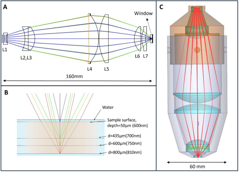
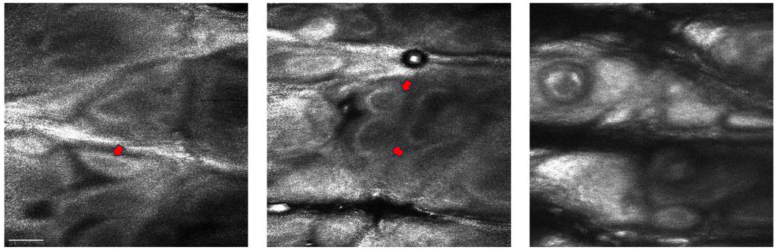
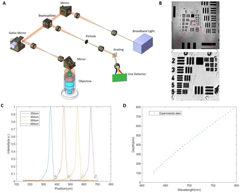
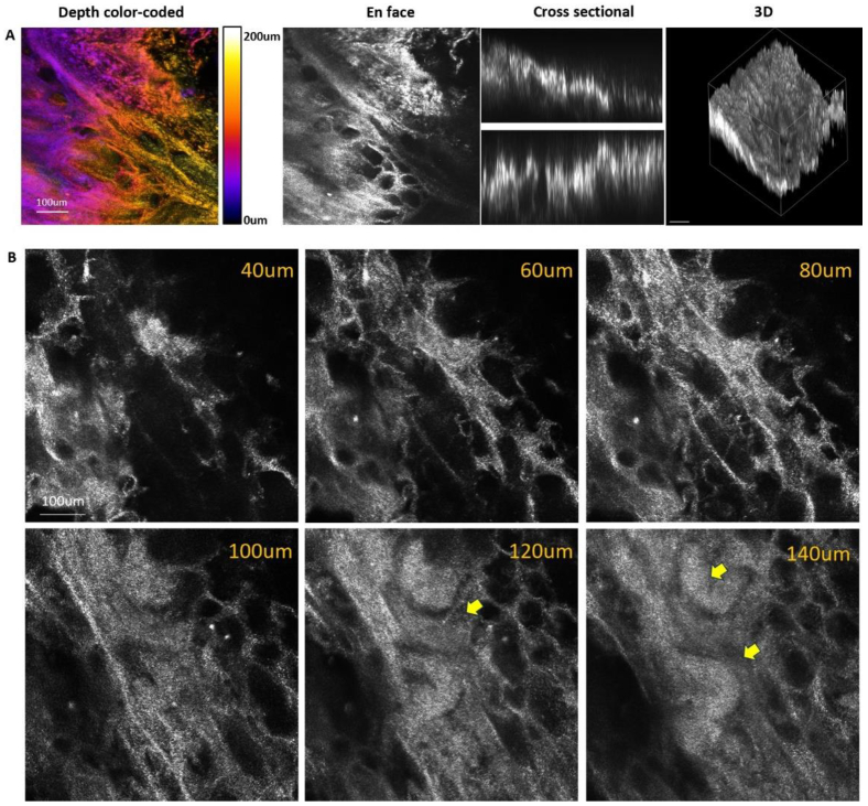
The limited focal shift of conventional achromatic objectives constrains the maximum imaging depth of chromatic confocal microscopes. To address this, we designed a hyperchromatic confocal microscope using diffractive optical elements, which was fabricated by single-point diamond turning (SPDT). This design takes advantage of the small Abbe number of diffractive optical elements to introduce a significant longitudinal chromatic shift. The resulting chromatic confocal microscope achieved a maximum imaging depth of 750 µm and a lateral resolution of 0.78 µm across a wavelength range of 600-810 nm. The system's imaging capabilities were demonstrated by capturing detailed images of biological samples, including cucumber seed cavities, pig kidney, and human forearm skin. These results confirmed the microscope's effectiveness in visualizing key cellular structures, underscoring its potential for high-resolution biological imaging.

摘要

传统消色差物镜有限的焦移限制了色差共聚焦显微镜的最大成像深度。为了解决这个问题,我们设计了一种使用衍射光学元件的超色差共聚焦显微镜,该显微镜通过单点金刚石车削(SPDT)制造。这种设计利用衍射光学元件较小的阿贝数来引入显著的纵向色差。由此产生的色差共聚焦显微镜在600-810nm的波长范围内实现了750μm的最大成像深度和0.78μm的横向分辨率。通过拍摄包括黄瓜种子腔、猪肾和人体前臂皮肤在内的生物样本的详细图像,展示了该系统的成像能力。这些结果证实了该显微镜在可视化关键细胞结构方面的有效性,突出了其在高分辨率生物成像方面的潜力。

https://cdn.ncbi.nlm.nih.gov/pmc/blobs/a8aa/11640585/77f0740fbb9f/boe-15-12-6834-g007.jpg
https://cdn.ncbi.nlm.nih.gov/pmc/blobs/a8aa/11640585/6ab7da02aa14/boe-15-12-6834-g001.jpg
https://cdn.ncbi.nlm.nih.gov/pmc/blobs/a8aa/11640585/8c591109c680/boe-15-12-6834-g002.jpg
https://cdn.ncbi.nlm.nih.gov/pmc/blobs/a8aa/11640585/d440dcba3856/boe-15-12-6834-g003.jpg
https://cdn.ncbi.nlm.nih.gov/pmc/blobs/a8aa/11640585/463c04af7e64/boe-15-12-6834-g004.jpg
https://cdn.ncbi.nlm.nih.gov/pmc/blobs/a8aa/11640585/5b13e6569e34/boe-15-12-6834-g005.jpg
https://cdn.ncbi.nlm.nih.gov/pmc/blobs/a8aa/11640585/35bf20ce6631/boe-15-12-6834-g006.jpg
https://cdn.ncbi.nlm.nih.gov/pmc/blobs/a8aa/11640585/77f0740fbb9f/boe-15-12-6834-g007.jpg

相似文献

1
Diffractive hyperchromatic objective for chromatic confocal microscopy.用于彩色共聚焦显微镜的衍射增色物镜。
Biomed Opt Express. 2024 Nov 14;15(12):6834-6844. doi: 10.1364/BOE.543322. eCollection 2024 Dec 1.
2
Chromatic confocal microscopy using staircase diffractive surface.使用阶梯衍射表面的彩色共聚焦显微镜。
Appl Opt. 2014 Aug 10;53(23):5123-30. doi: 10.1364/AO.53.005123.
3
Chromatic confocal microscopy for multi-depth imaging of epithelial tissue.用于上皮组织多深度成像的彩色共聚焦显微镜。
Biomed Opt Express. 2013 Apr 16;4(5):732-40. doi: 10.1364/BOE.4.000732. Print 2013 May 1.
4
Diffractive lenses for chromatic confocal imaging.用于彩色共焦成像的衍射透镜。
Appl Opt. 1997 Jul 10;36(20):4744-8. doi: 10.1364/ao.36.004744.
5
Hybrid diffractive-refractive lens for chromatic confocal measurement system.用于彩色共焦测量系统的混合衍射折射透镜。
Opt Express. 2024 May 20;32(11):20128-20135. doi: 10.1364/OE.525475.
6
Nontranslational three-dimensional profilometry by chromatic confocal microscopy with dynamically configurable micromirror scanning.采用动态可配置微镜扫描的彩色共聚焦显微镜进行非平移式三维轮廓测量。
Appl Opt. 2000 Jun 1;39(16):2605-13. doi: 10.1364/ao.39.002605.
7
Single-shot depth-section imaging through chromatic slit-scan confocal microscopy.通过彩色狭缝扫描共聚焦显微镜进行单次深度切片成像。
Appl Opt. 1998 Oct 1;37(28):6764-70. doi: 10.1364/ao.37.006764.
8
Ultra-broadband achromatic imaging with diffractive photon sieves.基于衍射光子筛的超宽带消色差成像
Sci Rep. 2016 Jun 22;6:28319. doi: 10.1038/srep28319.
9
Hyperchromatic lens doublets with an extremely small equivalent Abbe number employing diffractive elements and refractive materials with exceptional dispersion properties.采用具有极高色散特性的衍射元件和折射材料的、阿贝数极小的超色差胶合透镜对。
J Opt Soc Am A Opt Image Sci Vis. 2022 Nov 1;39(11):1992-2000. doi: 10.1364/JOSAA.473546.
10
Design, Analysis, and Manufacturing of Diffractive Achromatic Optical Systems.衍射消色差光学系统的设计、分析与制造
Micromachines (Basel). 2025 Mar 11;16(3):322. doi: 10.3390/mi16030322.

引用本文的文献

1
Label-free surface sectioning deep ultraviolet tissue imaging in multimodalities.多模态无标记表面切片深紫外组织成像
Biomed Opt Express. 2025 Jun 16;16(7):2756-2766. doi: 10.1364/BOE.565070. eCollection 2025 Jul 1.

本文引用的文献

1
Solvent-Free Silsesquioxane Self-Welding for 3D Printing Multi-Refractive Index Glass Objects.用于3D打印多折射率玻璃物体的无溶剂倍半硅氧烷自焊接
Adv Opt Mater. 2024 Aug 14;12(23). doi: 10.1002/adom.202400783. Epub 2024 Jun 5.
2
Bio-inspired Compact, High-resolution Snapshot Hyperspectral Imaging System with 3D Printed Glass Lightguide Array.具有3D打印玻璃光导阵列的受生物启发的紧凑型高分辨率快照高光谱成像系统
Adv Opt Mater. 2023 May 4;11(9). doi: 10.1002/adom.202300156. Epub 2023 Feb 28.
3
Experimental Studies on Fabricating Lenslet Array with Slow Tool Servo.
基于慢刀伺服加工小透镜阵列的实验研究
Micromachines (Basel). 2022 Sep 21;13(10):1564. doi: 10.3390/mi13101564.
4
High-Precision Printing of Complex Glass Imaging Optics with Precondensed Liquid Silica Resin.采用预缩合液态硅树脂对复杂玻璃成像光学器件进行高精度打印。
Adv Sci (Weinh). 2022 Jun;9(18):e2105595. doi: 10.1002/advs.202105595. Epub 2022 Apr 25.
5
Fiber-optic large-depth 3D chromatic confocal endomicroscopy.光纤大深度三维彩色共聚焦内镜显微镜
Biomed Opt Express. 2021 Dec 13;13(1):300-313. doi: 10.1364/BOE.442029. eCollection 2022 Jan 1.
6
DMD-based three-dimensional chromatic confocal microscopy.基于数字微镜器件的三维彩色共聚焦显微镜。
Appl Opt. 2020 May 10;59(14):4349-4356. doi: 10.1364/AO.386863.
7
Introduction to reflectance confocal microscopy and its use in clinical practice.反射式共聚焦显微镜简介及其在临床实践中的应用。
JAAD Case Rep. 2018 Nov 10;4(10):1014-1023. doi: 10.1016/j.jdcr.2018.09.019. eCollection 2018 Nov.
8
Chromatic confocal microscopy for multi-depth imaging of epithelial tissue.用于上皮组织多深度成像的彩色共聚焦显微镜。
Biomed Opt Express. 2013 Apr 16;4(5):732-40. doi: 10.1364/BOE.4.000732. Print 2013 May 1.
9
Theoretical analysis of confocal microscopy with microlenses.
Appl Opt. 1996 Jan 1;35(1):120-5. doi: 10.1364/AO.35.000120.
10
Three-dimensional image sensing by chromatic confocal microscopy.基于彩色共聚焦显微镜的三维图像传感
Appl Opt. 1994 Apr 1;33(10):1838-43. doi: 10.1364/AO.33.001838.