Abeysinghe Pevindu, Turner Natalie, Mitchell Murray D
Centre for Children's Health Research, Centre for Immunology and Infection Control, School of Biomedical Sciences, Faculty of Health, Queensland University of Technology, Brisbane, QLD 4101, Australia.
Centre for Genomics and Personalised Health, School of Biomedical Sciences, Faculty of Health, Queensland University of Technology, Kelvin Grove, QLD 4059, Australia.
Extracell Vesicles Circ Nucl Acids. 2024 Feb 29;5(1):119-137. doi: 10.20517/evcna.2023.55. eCollection 2024.
Analysis of miRNA (18-23nt) encapsulated in small extracellular vesicles (sEVs) (diameter ~30-200 nm) is critical in understanding the diagnostic and therapeutic value of sEV miRNA. However, various sEV enrichment techniques yield different quantities and qualities of sEV miRNA. Here, we compare the efficacy of three sEV isolation techniques in four combinations for miRNA next-generation sequencing.
Blood plasma from four Holstein-Friesian dairy cows () ( = 4) with similar genetic traits and physical characteristics were pooled to isolate sEV. Ultracentrifugation (UC) (100,000 × , 2 h at 4 °C), size-exclusion chromatography (SEC) and ultrafiltration (UF) were used to design four groups of sEV isolations (UC+SEC, SEC+UC, SEC+UF and UC+SEC+UF). sEV miRNAs were isolated using a combination of TRIzol, Chloroform and miRNeasy mini kit ( = 4/each), later sequenced utilizing Novaseq S1 platform (single-end 100 bp sequencing).
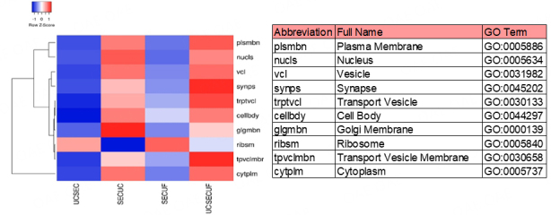
All four sEV methods yielded > 1,700 miRNAs and sEV miRNAs demonstrated a clear separation from control blood plasma circulating miRNA (PCA analysis). MiR-381-3p, miR-23-3p, and miR-18b-3p are among the 25 miRNAs unique to sEV, indicating potential sEV-specific miRNA markers. Further, those 25 miRNAs mostly regulate immune-related functions, indicating the value of sEV miRNA cargo in immunology.
The four sEV miRNA isolation methods employed in this study are valid techniques. The choice of method depends on the research question and study design. If purity is of concern, the UC+SEC method resulted in the best particles/µg protein ratio, which is often used as an indication of sample purity. These results could eventually establish sEV miRNAs as effective diagnostic and therapeutic tools of immunology.
分析包裹在小细胞外囊泡(直径约30 - 200 nm)中的微小RNA(18 - 23 nt)对于理解小细胞外囊泡微小RNA的诊断和治疗价值至关重要。然而,各种小细胞外囊泡富集技术产生的小细胞外囊泡微小RNA的数量和质量各不相同。在此,我们比较三种小细胞外囊泡分离技术的四种组合用于微小RNA下一代测序的效果。
将四头具有相似遗传特征和身体特征的荷斯坦 - 弗里生奶牛(n = 4)的血浆汇集以分离小细胞外囊泡。使用超速离心(100,000×g,4℃ 2小时)、尺寸排阻色谱法(SEC)和超滤法(UF)设计四组小细胞外囊泡分离方法(UC + SEC、SEC + UC、SEC + UF和UC + SEC + UF)。使用TRIzol、氯仿和miRNeasy迷你试剂盒组合分离小细胞外囊泡微小RNA(每组n = 4),随后利用Novaseq S1平台进行测序(单端100 bp测序)。
所有四种小细胞外囊泡方法均产生超过1700种微小RNA,并且小细胞外囊泡微小RNA与对照血浆循环微小RNA表现出明显分离(主成分分析)。MiR - 381 - 3p、miR - 23 - 3p和miR - 18b - 3p是小细胞外囊泡特有的25种微小RNA之一,表明存在潜在的小细胞外囊泡特异性微小RNA标志物。此外,这25种微小RNA大多调节免疫相关功能,表明小细胞外囊泡微小RNA货物在免疫学中的价值。
本研究采用的四种小细胞外囊泡微小RNA分离方法是有效的技术。方法的选择取决于研究问题和研究设计。如果关注纯度,UC + SEC方法产生的颗粒/μg蛋白质比率最佳,这通常用作样品纯度的指标。这些结果最终可能将小细胞外囊泡微小RNA确立为有效的免疫学诊断和治疗工具。