• 文献检索
  • 文档翻译
  • 深度研究
  • 学术资讯
  • Suppr Zotero 插件Zotero 插件
  • 邀请有礼
  • 套餐&价格
  • 历史记录
应用&插件
Suppr Zotero 插件Zotero 插件浏览器插件Mac 客户端Windows 客户端微信小程序
定价
高级版会员购买积分包购买API积分包
服务
文献检索文档翻译深度研究API 文档MCP 服务
关于我们
关于 Suppr公司介绍联系我们用户协议隐私条款
关注我们

Suppr 超能文献

核心技术专利:CN118964589B侵权必究
粤ICP备2023148730 号-1Suppr @ 2026

文献检索

告别复杂PubMed语法,用中文像聊天一样搜索,搜遍4000万医学文献。AI智能推荐,让科研检索更轻松。

立即免费搜索

文件翻译

保留排版,准确专业,支持PDF/Word/PPT等文件格式,支持 12+语言互译。

免费翻译文档

深度研究

AI帮你快速写综述,25分钟生成高质量综述,智能提取关键信息,辅助科研写作。

立即免费体验

纳米颗粒诱导酸蚀牙本质的仿生再矿化

Nanoparticles Induced Biomimetic Remineralization of Acid-Etched Dentin.

作者信息

Babu Devalla Venu, Ballullaya Srinidhi V, S Pushpa, Taufin Neha, Naveen Pilli Sai

机构信息

Dept. Conservative Dentistry and Endodontics, St.Joseph Dental College, Duggirala, Eluru, Andra Pradesh, India.

出版信息

J Dent (Shiraz). 2024 Dec 1;25(4):359-368. doi: 10.30476/dentjods.2024.98928.2117. eCollection 2024 Dec.

DOI:10.30476/dentjods.2024.98928.2117
PMID:39713104
原文链接:https://pmc.ncbi.nlm.nih.gov/articles/PMC11662173/
Abstract

STATEMENT OF THE PROBLEM

Dentin bonding with etch-and-rinse adhesives involves demineralizing the 5-8µm of the surface dentin to create micro space for resin infiltration. The presence of continuous fluid movement in dentin tubules and positive pulpal pressure prevents complete water replacement by resin monomers. This results in areas of demineralized dentin, which contain collagen fibers without resin infiltration. The exposed collage fibers are subjected to enzymatic degradation leading to less durable hybrid layer.

PURPOSE

The aim of this study was to evaluate the remineralizing effect of the nanoparticles on the resin dentin bonding interface.

MATERIALS AND METHOD

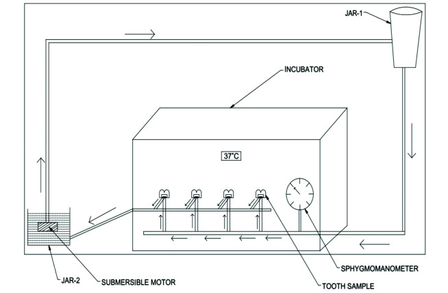
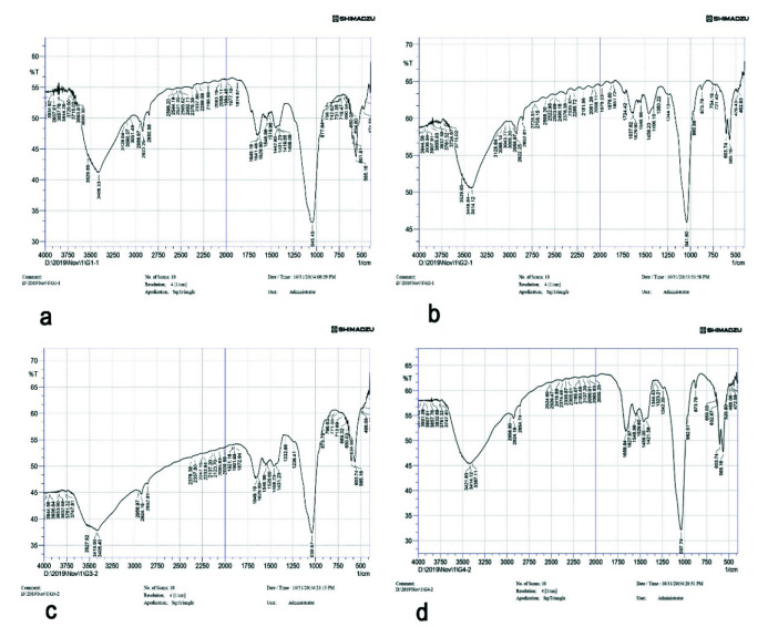
The three experimental remineralizing nanoparticles were characterized for their morphology, size, and composition. A total of 48 extracted non-carious human third molar teeth were sectioned at 2 mm below the cemento enamel junction. Class I cavity was prepared and the tooth samples were placed in an intra pulpal pressure simulation device. After etching of the prepared cavity, the samples were randomly divided into four groups (n=10) as follows: (1) control group(c) (n=10) (2) Nano-hydroxyapatite (nHAP) (n=10) (3) Chitosan-nanohydroxyapatite (Chi-nHAP) (n=10) (4) Mesoporous silica-hydrox-yapatite (MS-nHAP) (n=10). After 30 days remineralization period, the samples were evaluated for micro tensile bond strength, hybrid layer morphology, and mineral composition of the hybrid layer. The results were analyzed statistically by one-way ANOVA and Tukey's multiple post hoc tests.

RESULTS

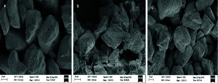
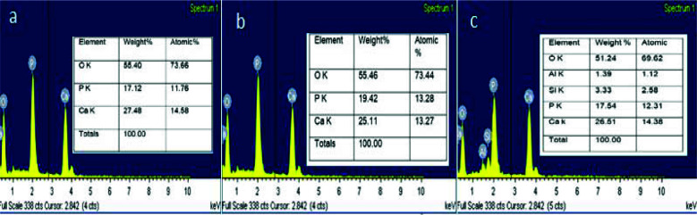
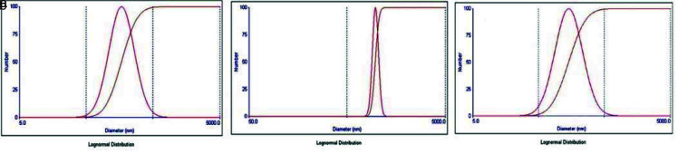
Scanning electron microscopic observation of nanoparticles revealed irregular particle shapes with calcium phosphate ratio of 1.60. The zeta analyzer showed a mean diameter of 161.0 nm, 323.0nm, 185.0nm for nHAP, Chi-nHAP, and MS-nHAP respectively. Post hoc Bonferroni test revealed significantly higher bond strength for nHAP, Chi-nHAP, and MS-nHAP when compared to control group. MS-nHAP resulted in the uniform deposition of apatite crystal on the surface without any evidence of dentinal tubules openings and had higher mineral to matrix ratio compared to other groups.

CONCLUSION

MS-nHAP nanoparticles can be considered as a reliable source of calcium and phosphate for biomimetic remineralization of hybrid layer. Application of nanoparticle remineralization precursors before application of dentin bonding agents results in remeralization of exposed collagen fibers thereby improving the clinical longevity of hybrid layer.

摘要

问题陈述

使用酸蚀冲洗粘结剂进行牙本质粘结时,需使表层5-8微米厚的牙本质脱矿,以形成树脂渗入的微空间。牙本质小管中持续的液体流动和牙髓内的正压,使得树脂单体无法完全取代水分。这导致脱矿牙本质区域存在未被树脂渗入的胶原纤维。暴露的胶原纤维会发生酶降解,致使混合层耐久性降低。

目的

本研究旨在评估纳米颗粒对树脂-牙本质粘结界面的再矿化作用。

材料与方法

对三种用于实验的再矿化纳米颗粒的形态、尺寸和成分进行了表征。选取48颗拔除的无龋人类第三磨牙,在牙骨质-釉质界下方2毫米处进行切片。制备I类洞型,并将牙齿样本置于牙髓内压力模拟装置中。对制备好的洞型进行酸蚀后,将样本随机分为四组(每组n = 10):(1)对照组(c)(n = 10);(2)纳米羟基磷灰石(nHAP)(n = 10);(3)壳聚糖-纳米羟基磷灰石(Chi-nHAP)(n = 10);(4)介孔二氧化硅-羟基磷灰石(MS-nHAP)(n = 10)。经过30天的再矿化期后,对样本进行微拉伸粘结强度、混合层形态以及混合层矿物成分的评估。结果采用单因素方差分析和Tukey多重事后检验进行统计学分析。

结果

纳米颗粒的扫描电子显微镜观察显示,颗粒形状不规则,磷酸钙比例为1.60。zeta分析仪显示,nHAP、Chi-nHAP和MS-nHAP的平均直径分别为161.0纳米、323.0纳米和185.0纳米。事后Bonferroni检验显示,与对照组相比,nHAP、Chi-nHAP和MS-nHAP的粘结强度显著更高。MS-nHAP使磷灰石晶体均匀沉积在表面,未发现牙本质小管开口的迹象,且与其他组相比,其矿物与基质的比例更高。

结论

MS-nHAP纳米颗粒可被视为混合层仿生再矿化的可靠钙磷来源。在应用牙本质粘结剂之前使用纳米颗粒再矿化前驱体,可使暴露的胶原纤维再矿化,从而提高混合层的临床耐久性。

https://cdn.ncbi.nlm.nih.gov/pmc/blobs/a958/11662173/38b6b8162861/JDS-25-359-g006.jpg
https://cdn.ncbi.nlm.nih.gov/pmc/blobs/a958/11662173/20eb708ec1bd/JDS-25-359-g001.jpg
https://cdn.ncbi.nlm.nih.gov/pmc/blobs/a958/11662173/1d19317c4150/JDS-25-359-g002.jpg
https://cdn.ncbi.nlm.nih.gov/pmc/blobs/a958/11662173/66c42a9c6734/JDS-25-359-g003.jpg
https://cdn.ncbi.nlm.nih.gov/pmc/blobs/a958/11662173/19649b742ccb/JDS-25-359-g004.jpg
https://cdn.ncbi.nlm.nih.gov/pmc/blobs/a958/11662173/d5ed503069e6/JDS-25-359-g005.jpg
https://cdn.ncbi.nlm.nih.gov/pmc/blobs/a958/11662173/38b6b8162861/JDS-25-359-g006.jpg

相似文献

1
Nanoparticles Induced Biomimetic Remineralization of Acid-Etched Dentin.纳米颗粒诱导酸蚀牙本质的仿生再矿化
J Dent (Shiraz). 2024 Dec 1;25(4):359-368. doi: 10.30476/dentjods.2024.98928.2117. eCollection 2024 Dec.
2
Zinc-modified nanopolymers improve the quality of resin-dentin bonded interfaces.锌改性纳米聚合物可改善树脂-牙本质粘结界面的质量。
Clin Oral Investig. 2016 Dec;20(9):2411-2420. doi: 10.1007/s00784-016-1738-y. Epub 2016 Jan 30.
3
The influence of neutral MDP-Na salt on dentin bond performance and remineralization potential of etch-&-rinse adhesive.中性 MDP-Na 盐对酸蚀-冲洗型黏结剂牙本质黏结性能和再矿化潜力的影响。
BMC Oral Health. 2024 Aug 24;24(1):997. doi: 10.1186/s12903-024-04756-y.
4
Oyster shell-derived nano-hydroxyapatite and proanthocyanidin pretreatment on dentinal tubule occlusion and permeability before and after acid challenge-an in vitro study.牡蛎壳衍生纳米羟基磷灰石和原花青素预处理对酸蚀前后牙本质小管闭塞和通透性的影响:一项体外研究。
J Mater Sci Mater Med. 2023 Apr 10;34(4):17. doi: 10.1007/s10856-023-06724-4.
5
Influence of nano-hydroxyapatite containing desensitizing toothpastes on the sealing ability of dentinal tubules and bonding performance of self-etch adhesives.含纳米羟磷灰石脱敏牙膏对牙本质小管封闭能力和自酸蚀粘结剂粘结性能的影响。
J Mech Behav Biomed Mater. 2019 Mar;91:38-44. doi: 10.1016/j.jmbbm.2018.11.021. Epub 2018 Nov 28.
6
Evaluation of the effect of remineralization with strontium-doped nanohydroxyapatite with noncollagenous protein analog: Chitosan on the shear bond strength of resin composite to dentin - An study.用含非胶原蛋白类似物壳聚糖的掺锶纳米羟基磷灰石再矿化对树脂复合材料与牙本质剪切粘结强度影响的评估——一项研究。
J Conserv Dent. 2022 Nov-Dec;25(6):648-653. doi: 10.4103/jcd.jcd_285_22. Epub 2022 Oct 13.
7
Influence of phosphoproteins' biomimetic analogs on remineralization of mineral-depleted resin-dentin interfaces created with ion-releasing resin-based systems.磷蛋白仿生类似物对使用离子释放型树脂基系统创建的矿物质耗尽的树脂-牙本质界面再矿化的影响。
Dent Mater. 2015 Jul;31(7):759-77. doi: 10.1016/j.dental.2015.03.013. Epub 2015 Apr 23.
8
Bonding performance of experimental bioactive/biomimetic self-etch adhesives doped with calcium-phosphate fillers and biomimetic analogs of phosphoproteins.实验性生物活性/仿生自酸蚀黏合剂掺杂钙磷填料和磷蛋白仿生模拟物的黏结性能。
J Dent. 2016 Sep;52:79-86. doi: 10.1016/j.jdent.2016.07.016. Epub 2016 Jul 27.
9
Biomimetic remineralization of resin-bonded acid-etched dentin.树脂粘结酸蚀牙本质的仿生再矿化
J Dent Res. 2009 Aug;88(8):719-24. doi: 10.1177/0022034509341826.
10
The use of bioactive particles and biomimetic analogues for increasing the longevity of resin-dentin interfaces: A literature review.生物活性颗粒和仿生类似物在增加树脂-牙本质界面寿命中的应用:文献综述。
Dent Mater J. 2020 Jan 31;39(1):62-68. doi: 10.4012/dmj.2019-293. Epub 2019 Nov 14.

本文引用的文献

1
The effect of silicate ions on proliferation, osteogenic differentiation and cell signalling pathways (WNT and SHH) of bone marrow stromal cells.硅酸根离子对骨髓基质细胞增殖、成骨分化及细胞信号通路(WNT和SHH)的影响
Biomater Sci. 2013 Apr 5;1(4):379-392. doi: 10.1039/c2bm00108j. Epub 2012 Dec 12.
2
Effect of different sizes of bioactive glass-coated mesoporous silica nanoparticles on dentinal tubule occlusion and mineralization.不同粒径的载有活性玻璃的介孔硅纳米颗粒对牙本质小管封闭和矿化的影响。
Clin Oral Investig. 2019 May;23(5):2129-2141. doi: 10.1007/s00784-018-2658-9. Epub 2018 Sep 30.
3
Biodegradable mesoporous delivery system for biomineralization precursors.
用于生物矿化前体的可生物降解介孔递送系统。
Int J Nanomedicine. 2017 Jan 25;12:839-854. doi: 10.2147/IJN.S128792. eCollection 2017.
4
Translation of a solution-based biomineralization concept into a carrier-based delivery system via the use of expanded-pore mesoporous silica.通过使用扩孔介孔二氧化硅,将基于溶液的生物矿化概念转化为基于载体的递送系统。
Acta Biomater. 2016 Feb;31:378-387. doi: 10.1016/j.actbio.2015.11.062. Epub 2015 Dec 2.
5
Biomimetic remineralization of demineralized dentine using scaffold of CMC/ACP nanocomplexes in an in vitro tooth model of deep caries.在深龋体外牙齿模型中使用羧甲基纤维素/无定形磷酸钙纳米复合物支架对脱矿牙本质进行仿生再矿化。
PLoS One. 2015 Jan 14;10(1):e0116553. doi: 10.1371/journal.pone.0116553. eCollection 2015.
6
A mesoporous silica biomaterial for dental biomimetic crystallization.一种用于牙齿仿生结晶的介孔硅生物材料。
ACS Nano. 2014 Dec 23;8(12):12502-13. doi: 10.1021/nn5053487. Epub 2014 Dec 11.
7
Remineralization potential of fully demineralized dentin infiltrated with silica and hydroxyapatite nanoparticles.用二氧化硅和羟基磷灰石纳米颗粒渗透的完全脱矿牙本质的再矿化潜力。
Dent Mater. 2014 Mar;30(3):249-62. doi: 10.1016/j.dental.2013.11.014. Epub 2014 Jan 17.
8
Influence of different intrapulpal pressure simulation liquids on the microtensile bond strength of adhesive systems to dentin.不同牙髓内压力模拟液对牙本质黏结系统微拉伸黏结强度的影响。
J Adhes Dent. 2013 Dec;15(6):519-26. doi: 10.3290/j.jad.a29719.
9
Infiltration of demineralized dentin with silica and hydroxyapatite nanoparticles.脱矿牙本质内的二氧化硅和羟基磷灰石纳米粒子渗透。
Dent Mater. 2012 Sep;28(9):1012-23. doi: 10.1016/j.dental.2012.05.007. Epub 2012 Jun 19.
10
Bonding effectiveness of a new 'multi-mode' adhesive to enamel and dentine.新型“多模式”黏合剂对牙釉质和牙本质黏结效果的评价。
J Dent. 2012 Jun;40(6):475-84. doi: 10.1016/j.jdent.2012.02.012. Epub 2012 Feb 28.