Wang Fang, Han Hailong, Wang Caifang, Wang Jingfei, Peng Yanni, Chen Ye, He Yaohui, Deng Zhouyang, Li Fang, Rong Yikang, Wang Danling, Liu Wen, Chen Hualan, Zhang Zhuohua
Department of Neurosciences, Hengyang Medical School, University of South China, Hengyang, 421009, China.
Institute of Molecular Precision Medicine and Hunan Provincial Key Laboratory of Molecular Precision Medicine, Xiangya Hospital, Central South University, Changsha, 410078, China.
Transl Neurodegener. 2024 Dec 27;13(1):68. doi: 10.1186/s40035-024-00458-1.
Neurological complications are a significant concern of Coronavirus Disease 2019 (COVID-19). However, the pathogenic mechanism of neurological symptoms associated with severe acute respiratory syndrome coronavirus 2 (SARS-CoV-2) infection is poorly understood.
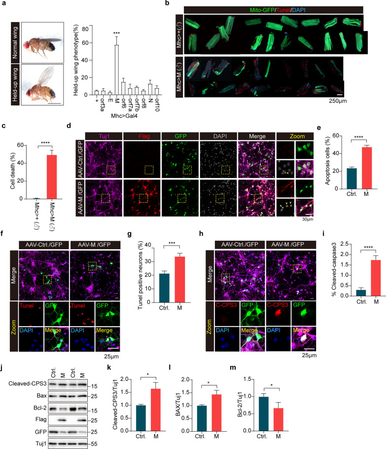
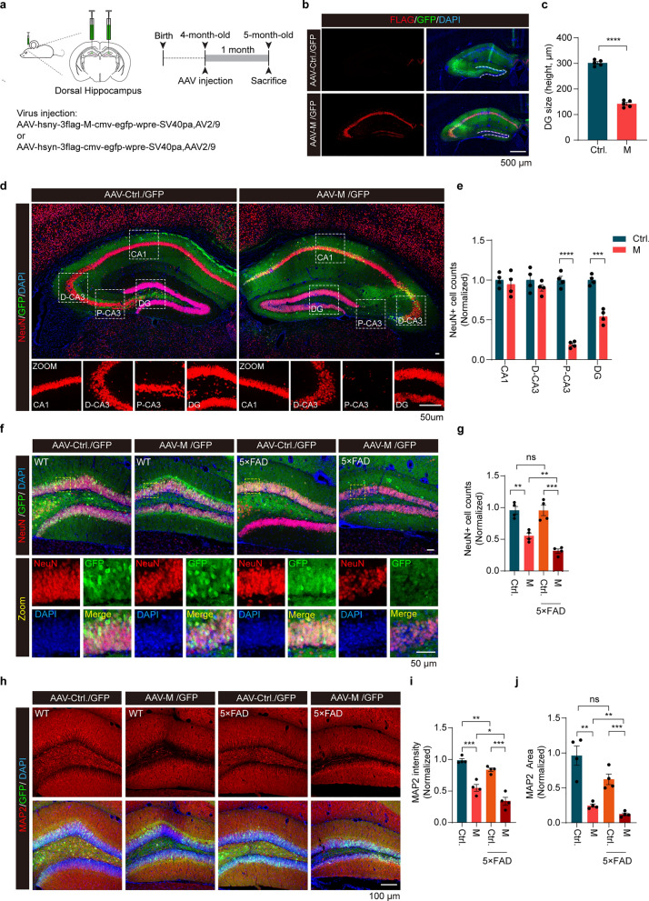
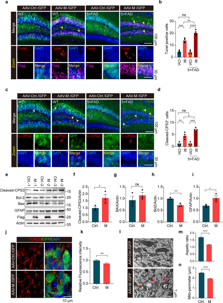
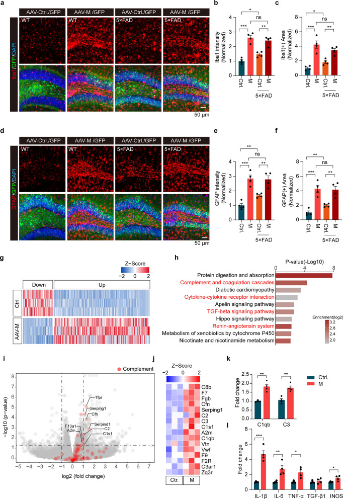
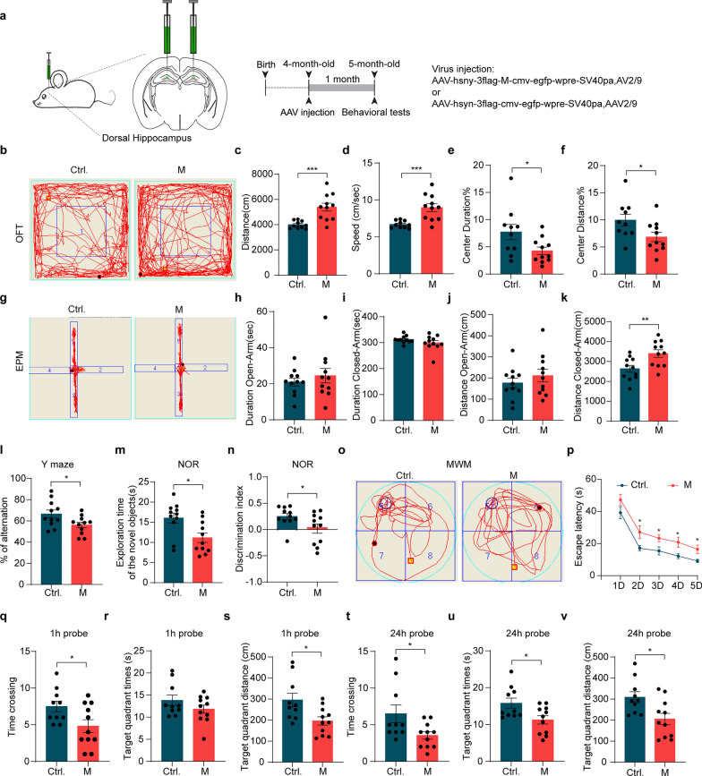
We used Drosophila as a model to systematically analyze SARS-CoV-2 genes encoding structural and accessory proteins and identified the membrane protein (M) that disrupted mitochondrial functions in vivo. The M protein was stereotaxically injected to further assess its effects in the brains of wild-type (WT) and 5 × FAD mice. Omics technologies, including RNA sequencing and interactome analysis, were performed to explore the mechanisms of the effects of M protein both in vitro and in vivo.
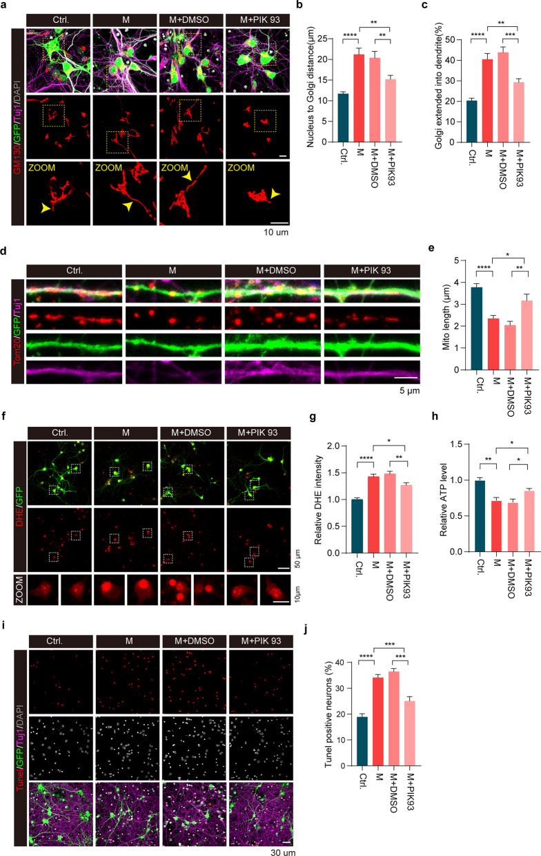
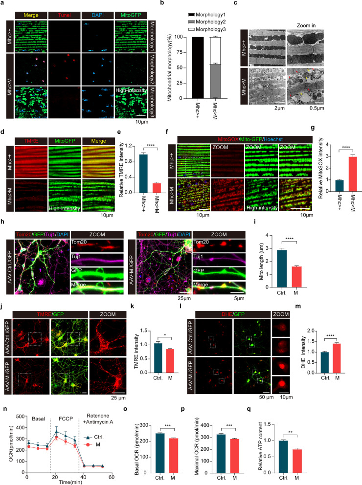
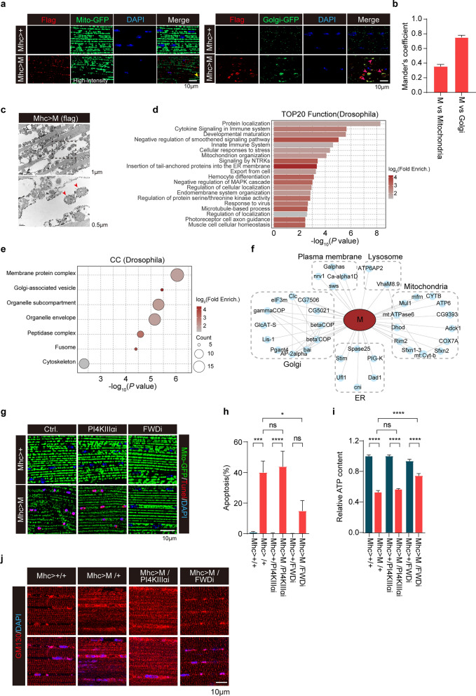
Systematic analysis of SARS-CoV-2 structural and accessory proteins in Drosophila identified that the M protein induces mitochondrial fragmentation and dysfunction, leading to reduced ATP production, ROS overproduction, and eventually cell death in the indirect flight muscles. In WT mice, M caused hippocampal atrophy, neural apoptosis, glial activation, and mitochondrial damage. These changes were further aggravated in 5 × FAD mice. M was localized to the Golgi apparatus and genetically interacted with four wheel drive (FWD, a Drosophila homolog of mammalian PI4KIIIβ) to regulate Golgi functions in flies. Fwd RNAi, but not PI4KIIIα RNAi, reversed the M-induced Golgi abnormality, mitochondrial fragmentation, and ATP reduction. Inhibition of PI4KIIIβ activity suppressed the M-induced neuronal cell death. Therefore, M induced mitochondrial fragmentation and apoptosis likely through disruption of Golgi-derived PI(4)P-containing vesicles.
M disturbs the distribution and function of Golgi, leading to mitochondrial abnormality and eventually neurodegeneration via a PI4KIIIβ-mediated mechanism. This study reveals a potential mechanism for COVID-19 neurological symptoms and opens a new avenue for development of therapeutic strategies targeting SARS-CoV-2 M or mitochondria.
神经并发症是2019冠状病毒病(COVID-19)的一个重要问题。然而,与严重急性呼吸综合征冠状病毒2(SARS-CoV-2)感染相关的神经症状的致病机制尚不清楚。
我们以果蝇为模型,系统分析了SARS-CoV-2编码结构蛋白和辅助蛋白的基因,并鉴定出在体内破坏线粒体功能的膜蛋白(M)。将M蛋白立体定向注射到野生型(WT)和5×FAD小鼠的大脑中,以进一步评估其作用。采用包括RNA测序和相互作用组分析在内的组学技术,探索M蛋白在体外和体内发挥作用的机制。
对果蝇体内SARS-CoV-2结构蛋白和辅助蛋白的系统分析表明,M蛋白诱导线粒体碎片化和功能障碍,导致间接飞行肌中ATP生成减少、活性氧过度产生,最终导致细胞死亡。在野生型小鼠中,M蛋白导致海马萎缩、神经细胞凋亡、胶质细胞活化和线粒体损伤。在5×FAD小鼠中,这些变化进一步加剧。M蛋白定位于高尔基体,并与四轮驱动蛋白(FWD,哺乳动物PI4KIIIβ的果蝇同源物)发生遗传相互作用,以调节果蝇体内的高尔基体功能。Fwd RNA干扰而非PI4KIIIα RNA干扰可逆转M蛋白诱导的高尔基体异常、线粒体碎片化和ATP减少。抑制PI4KIIIβ活性可抑制M蛋白诱导的神经元细胞死亡。因此,M蛋白可能通过破坏高尔基体衍生的含PI(4)P囊泡诱导线粒体碎片化和细胞凋亡。
M蛋白扰乱高尔基体的分布和功能,通过PI4KIIIβ介导的机制导致线粒体异常,最终引发神经退行性变。本研究揭示了COVID-19神经症状的潜在机制,并为开发针对SARS-CoV-2 M蛋白或线粒体的治疗策略开辟了新途径。