Aasa Maja, Lindquist David, Ottander Ulrika, Strandberg Sara N
Department of Diagnostics and Intervention, Diagnostic Radiology, Umeå University, Umeå, Sweden.
Department of Clinical Sciences, Professional Development, Umeå University, Umeå, Sweden.
EJNMMI Rep. 2025 Jan 10;9(1):3. doi: 10.1186/s41824-024-00236-2.
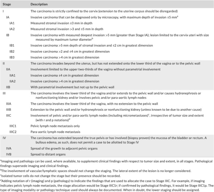
In uterine cervical cancer (UCC), tumour staging is performed according to the 2018 International Federation of Gynecology and Obstetrics (FIGO) system, where imaging is incorporated, or the more generic Tumour Node Metastasis (TNM) classification. With the technical development in diagnostic imaging, continuous prospective evaluation of the different imaging methods contributing to stage determination is warranted. The aims of this interim study were to (1) evaluate the performance of radiological FIGO (rFIGO) and T staging (rT) with 2-fluorine-18-fluoro-deoxy-glucose (2[18F]-FDG)-positron emission tomography with computed tomography (PET/CT) and with magnetic resonance imaging (PET/MRI), compared to clinical FIGO (cFIGO) and T (cT) staging based on clinical examination and conventional imaging, in treatment-naïve UCC, and to (2) identify possible MRI biomarkers for early treatment response after radiotherapy.
Ten consecutive patients with newly diagnosed UCC from the prospective PRODIGYN (Prognostic and Diagnostic Added Value of Medical Imaging in Staging and Treatment Planning of Gynecological Cancer) study (ethical approval number 2022-04207-01; NCT05855941) were included. Study participants underwent 2[18F]FDG-PET/CT and -PET/MRI, and an additional MRI one week after radiotherapy. Agreement between rFIGO and cFIGO was analysed using Cohen's kappa. Differences in rFIGO between 2[18F]FDG-PET/CT and -PET/MRI were evaluated with Wilcoxon signed ranks test, and added value of rFIGO for metastasis assessment was demonstrated with descriptive statistics.
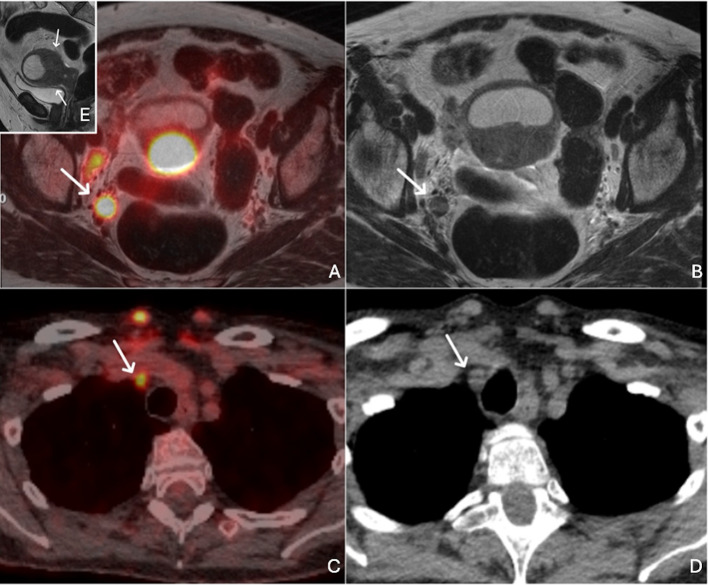
In 2/10 patients, a higher stage was obtained with rFIGO compared to cFIGO, where presence of metastases led to upstaging. In 3/10, rFIGO was lower than cFIGO, and in 5/10 rFIGO and cFIGO were similar. Degree of agreement between rFIGO and cFIGO was poor, (κ = 0.091, p < 0.005) with 2[18F]FDG-PET/CT and (κ = - 0.010, p > 0.05) with FDG/PET/MRI). There was no significant difference between 2[18F]FDG-PET/CT and -PET/MRI for rFIGO (p = 0.18), or rT stage assessment (p = 0.32). MRI-derived tumour volume and apparent diffusion coefficient (ADC) were most affected on MRI one week after radiotherapy.
Our results indicate that there is an added value of rFIGO staging with 2[18F]FDG-PET/CT and -PET/MRI compared to clinical examination and conventional radiology, for metastasis assessment in treatment-naïve UCC. In early treatment response evaluation with MRI, ADC and tumour volume may be predictive parameters of interest in future prognostic analyses.
Clinical Trials, NCT05855941. Registered 02 May 2023, https://clinicaltrials.gov/study/NCT05855941?term=NCT05855941&rank=1 .
在子宫颈癌(UCC)中,肿瘤分期是根据2018年国际妇产科联盟(FIGO)系统进行的,该系统纳入了影像学检查,或者采用更通用的肿瘤-淋巴结-转移(TNM)分类。随着诊断成像技术的发展,有必要对有助于分期确定的不同成像方法进行持续的前瞻性评估。这项中期研究的目的是:(1)在未经治疗的UCC中,将基于2-氟-18-氟脱氧葡萄糖(2[¹⁸F]-FDG)正电子发射断层扫描与计算机断层扫描(PET/CT)以及磁共振成像(PET/MRI)的放射学FIGO(rFIGO)和T分期(rT)的性能,与基于临床检查和传统成像的临床FIGO(cFIGO)和T(cT)分期进行比较;(2)识别放疗后早期治疗反应的可能MRI生物标志物。
纳入了前瞻性PRODIGYN(医学成像在妇科癌症分期和治疗计划中的预后和诊断附加值)研究中连续的10例新诊断的UCC患者(伦理批准号2022-04207-01;NCT05855941)。研究参与者接受了2[¹⁸F]FDG-PET/CT和PET/MRI检查,并在放疗一周后额外进行了一次MRI检查。使用Cohen's kappa分析rFIGO和cFIGO之间的一致性。采用Wilcoxon符号秩检验评估2[¹⁸F]FDG-PET/CT和PET/MRI之间rFIGO的差异,并用描述性统计展示rFIGO在转移评估中的附加值。
在10例患者中的2例中,与cFIGO相比,rFIGO获得了更高的分期,转移的存在导致了分期上调。在10例中的3例中,rFIGO低于cFIGO,在10例中的5例中,rFIGO和cFIGO相似。rFIGO和cFIGO之间的一致性程度较差,2[¹⁸F]FDG-PET/CT时(κ = 0.091,p < 0.005),FDG/PET/MRI时(κ = -0.010,p > 0.05)。2[¹⁸F]FDG-PET/CT和PET/MRI在rFIGO(p = 0.18)或rT分期评估(p = 0.32)方面没有显著差异。放疗一周后的MRI上,MRI衍生的肿瘤体积和表观扩散系数(ADC)受影响最大。
我们的结果表明,与临床检查和传统放射学相比,2[¹⁸F]FDG-PET/CT和PET/MRI的rFIGO分期在未经治疗的UCC转移评估中具有附加值。在MRI评估早期治疗反应时,ADC和肿瘤体积可能是未来预后分析中感兴趣的预测参数。
临床试验,NCT05855941。于2023年5月2日注册,https://clinicaltrials.gov/study/NCT05855941?term=NCT05855941&rank=1 。