• 文献检索
  • 文档翻译
  • 深度研究
  • 学术资讯
  • Suppr Zotero 插件Zotero 插件
  • 邀请有礼
  • 套餐&价格
  • 历史记录
应用&插件
Suppr Zotero 插件Zotero 插件浏览器插件Mac 客户端Windows 客户端微信小程序
定价
高级版会员购买积分包购买API积分包
服务
文献检索文档翻译深度研究API 文档MCP 服务
关于我们
关于 Suppr公司介绍联系我们用户协议隐私条款
关注我们

Suppr 超能文献

核心技术专利:CN118964589B侵权必究
粤ICP备2023148730 号-1Suppr @ 2026

文献检索

告别复杂PubMed语法,用中文像聊天一样搜索,搜遍4000万医学文献。AI智能推荐,让科研检索更轻松。

立即免费搜索

文件翻译

保留排版,准确专业,支持PDF/Word/PPT等文件格式,支持 12+语言互译。

免费翻译文档

深度研究

AI帮你快速写综述,25分钟生成高质量综述,智能提取关键信息,辅助科研写作。

立即免费体验

18F-FDG PET/MRI对原发性食管癌/胃食管癌进行分期的初步经验。

Initial experience in staging primary oesophageal/gastro-oesophageal cancer with 18F-FDG PET/MRI.

作者信息

Sharkey Amy R, Sah Bert-Ram, Withey Samuel J, Bhuva Shaheel, Neji Radhouene, Jeljeli Sami, Green Adrian, Cook Gary J R, Goh Vicky

机构信息

Department of Cancer Imaging, School of Biomedical Engineering and Imaging Sciences, King's College London, London, UK.

Department of Radiology, Guy's and St Thomas' NHS Foundation Trust, London, UK.

出版信息

Eur J Hybrid Imaging. 2021 Dec 13;5(1):23. doi: 10.1186/s41824-021-00117-y.

DOI:10.1186/s41824-021-00117-y
PMID:34897589
原文链接:https://pmc.ncbi.nlm.nih.gov/articles/PMC8666393/
Abstract

BACKGROUND

F-fluorodeoxyglucose positron emission tomography/magnetic resonance imaging (18F-FDG PET/MRI) may improve cancer staging by combining sensitive cancer detection with high-contrast resolution and detail. We compared the diagnostic performance of 18F-FDG PET/MRI to F-fluorodeoxyglucose positron emission tomography/computed tomography (18F-FDG PET/CT) for staging oesophageal/gastro-oesophageal cancer. Following ethical approval and informed consent, participants with newly diagnosed primary oesophageal/gastro-oesophageal cancer were enrolled. Exclusions included prior/concurrent malignancy. Following 324 ± 28 MBq 18F-FDG administration and 60-min uptake, PET/CT was performed, immediately followed by integrated PET/MRI from skull base to mid-thigh. PET/CT was interpreted by two dual-accredited nuclear medicine physicians and PET/MRI by a dual-accredited nuclear medicine physician/radiologist and cancer radiologist in consensus. Per-participant staging was compared with the tumour board consensus staging using the McNemar test, with statistical significance at 5%.

RESULTS

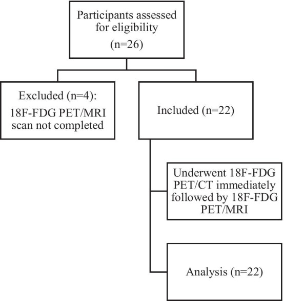
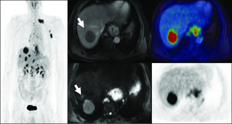
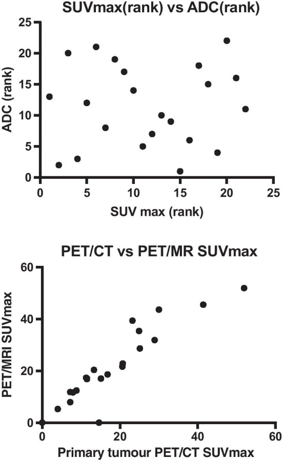
Out of 26 participants, 22 (20 males; mean ± SD age 68.8 ± 8.7 years) completed 18F-FDG PET/CT and PET/MRI. Compared to the tumour board, the primary tumour was staged concordantly in 55% (12/22) with PET/MRI and 36% (8/22) with PET/CT; the nodal stage was concordant in 45% (10/22) with PET/MRI and 50% (11/22) with PET/CT. There was no statistical difference in PET/CT and PET/MRI staging performance (p > 0.05, for T and N staging). The staging of distant metastases was concordant with the tumour board in 95% (21/22) with both PET/MRI and PET/CT. Of participants with distant metastatic disease, PET/MRI detected additional metastases in 30% (3/10).

CONCLUSION

In this preliminary study, compared to 18F-FDG PET/CT, 18F-FDG PET/MRI showed non-significant higher concordance with T-staging, but no difference with N or M-staging. Additional metastases detected by 18F-FDG PET/MRI may be of additive clinical value.

摘要

背景

氟脱氧葡萄糖正电子发射断层扫描/磁共振成像(18F-FDG PET/MRI)可通过将灵敏的癌症检测与高对比度分辨率及细节相结合来改善癌症分期。我们比较了18F-FDG PET/MRI与氟脱氧葡萄糖正电子发射断层扫描/计算机断层扫描(18F-FDG PET/CT)对食管癌/胃食管癌进行分期的诊断性能。在获得伦理批准并取得知情同意后,纳入新诊断的原发性食管癌/胃食管癌患者。排除标准包括既往/同时存在的恶性肿瘤。静脉注射324±28 MBq 18F-FDG并经过60分钟摄取后,进行PET/CT检查,随后立即进行从头颅底部至大腿中部的PET/MRI联合检查。PET/CT由两名具有双重资质的核医学医师解读,PET/MRI由一名具有双重资质的核医学医师/放射科医师和一名癌症放射科医师共同解读并达成共识。使用McNemar检验将每位参与者的分期与肿瘤委员会的共识分期进行比较,以5%作为统计学显著性水平。

结果

26名参与者中,22名(20名男性;平均±标准差年龄68.8±8.7岁)完成了18F-FDG PET/CT和PET/MRI检查。与肿瘤委员会的分期相比,PET/MRI对原发肿瘤分期的一致性为55%(12/22),PET/CT为36%(8/22);PET/MRI对区域淋巴结分期的一致性为45%(10/22),PET/CT为50%(11/22)。PET/CT和PET/MRI的分期性能无统计学差异(T分期和N分期的p>0.05)。PET/MRI和PET/CT对远处转移分期与肿瘤委员会的一致性均为95%(21/22)。在有远处转移的参与者中,PET/MRI额外检测到30%(3/10)的转移灶。

结论

在这项初步研究中,与18F-FDG PET/CT相比,18F-FDG PET/MRI在T分期方面显示出非显著性更高的一致性,但在N分期或M分期方面无差异。18F-FDG PET/MRI检测到的额外转移灶可能具有附加临床价值。

https://cdn.ncbi.nlm.nih.gov/pmc/blobs/0dc2/8666393/eedfaac71a6c/41824_2021_117_Fig4_HTML.jpg
https://cdn.ncbi.nlm.nih.gov/pmc/blobs/0dc2/8666393/c0d485cb8c47/41824_2021_117_Fig1_HTML.jpg
https://cdn.ncbi.nlm.nih.gov/pmc/blobs/0dc2/8666393/40f0734ee777/41824_2021_117_Fig2_HTML.jpg
https://cdn.ncbi.nlm.nih.gov/pmc/blobs/0dc2/8666393/cd7a99744dc6/41824_2021_117_Fig3_HTML.jpg
https://cdn.ncbi.nlm.nih.gov/pmc/blobs/0dc2/8666393/eedfaac71a6c/41824_2021_117_Fig4_HTML.jpg

相似文献

1
Initial experience in staging primary oesophageal/gastro-oesophageal cancer with 18F-FDG PET/MRI.18F-FDG PET/MRI对原发性食管癌/胃食管癌进行分期的初步经验。
Eur J Hybrid Imaging. 2021 Dec 13;5(1):23. doi: 10.1186/s41824-021-00117-y.
2
More advantages in detecting bone and soft tissue metastases from prostate cancer using F-PSMA PET/CT.使用F-PSMA PET/CT检测前列腺癌骨和软组织转移方面有更多优势。
Hell J Nucl Med. 2019 Jan-Apr;22(1):6-9. doi: 10.1967/s002449910952. Epub 2019 Mar 7.
3
What Is the Comparative Ability of 18F-FDG PET/CT, 99mTc-MDP Skeletal Scintigraphy, and Whole-body MRI as a Staging Investigation to Detect Skeletal Metastases in Patients with Osteosarcoma and Ewing Sarcoma?18F-FDG PET/CT、99mTc-MDP 骨显像和全身 MRI 作为分期检查在检测骨肉瘤和尤文肉瘤患者骨转移中的比较能力如何?
Clin Orthop Relat Res. 2021 Aug 1;479(8):1768-1779. doi: 10.1097/CORR.0000000000001681.
4
F-FDG-PET/MRI in preoperative staging of oesophageal and gastroesophageal junctional cancer.氟代脱氧葡萄糖正电子发射断层扫描/磁共振成像在食管和胃食管交界处癌术前分期中的应用。
Clin Radiol. 2019 Sep;74(9):718-725. doi: 10.1016/j.crad.2019.05.016. Epub 2019 Jun 18.
5
Thoracic staging in lung cancer: prospective comparison of 18F-FDG PET/MR imaging and 18F-FDG PET/CT.肺癌的胸部分期:18F-FDG PET/MR 成像与 18F-FDG PET/CT 的前瞻性比较。
J Nucl Med. 2014 Mar;55(3):373-8. doi: 10.2967/jnumed.113.129825. Epub 2014 Feb 6.
6
Dual-time point F-FDG PET/CT for the staging of oesophageal cancer: the best diagnostic performance by retention index for N-staging in non-calcified lymph nodes.双时相 F-FDG PET/CT 用于食管癌分期:非钙化淋巴结中保留指数对 N 分期的最佳诊断性能。
Eur J Nucl Med Mol Imaging. 2018 Jul;45(8):1317-1328. doi: 10.1007/s00259-018-3981-8. Epub 2018 Mar 3.
7
Primary staging with 2[18F]-FDG-PET/CT and -PET/MRI and radiotherapy response evaluation with MRI in uterine cervical cancer: an interim analysis of a prospective clinical trial.2-[18F]-FDG-PET/CT和PET/MRI用于子宫颈癌的初始分期以及MRI用于放疗反应评估:一项前瞻性临床试验的中期分析
EJNMMI Rep. 2025 Jan 10;9(1):3. doi: 10.1186/s41824-024-00236-2.
8
Breast cancer staging in a single session: whole-body PET/CT mammography.单次检查完成乳腺癌分期:全身PET/CT乳腺造影
J Nucl Med. 2008 Aug;49(8):1215-22. doi: 10.2967/jnumed.108.052050. Epub 2008 Jul 16.
9
Preoperative lymph-node staging of invasive urothelial bladder cancer with 18F-fluorodeoxyglucose positron emission tomography/computed axial tomography and magnetic resonance imaging: correlation with histopathology.18F-氟脱氧葡萄糖正电子发射断层扫描/计算机断层扫描和磁共振成像对浸润性膀胱尿路上皮癌的术前淋巴结分期:与组织病理学的相关性
Scand J Urol Nephrol. 2011 Mar;45(2):122-8. doi: 10.3109/00365599.2010.544672. Epub 2011 Jan 13.
10
Comparison of whole-body 18F-FDG PET/CT and PET/MRI for distant metastases in patients with malignant tumors: a meta-analysis.全身 18F-FDG PET/CT 和 PET/MRI 对恶性肿瘤患者远处转移的比较:一项荟萃分析。
BMC Cancer. 2023 Jan 10;23(1):37. doi: 10.1186/s12885-022-10493-8.

引用本文的文献

1
The prognostic value of positron emission tomography/magnetic resonance imaging in predicting survival in patients with adenocarcinoma of the esophagogastric junction.正电子发射断层扫描/磁共振成像在预测食管胃交界腺癌患者生存情况中的预后价值
Ann Nucl Med. 2025 May 17. doi: 10.1007/s12149-025-02058-z.
2
PET/MRI is superior to PET/CT in detecting oesophago and gastric carcinomas: a meta-analysis.PET/MRI在检测食管癌和胃癌方面优于PET/CT:一项荟萃分析。
Cancer Imaging. 2025 Apr 7;25(1):50. doi: 10.1186/s40644-025-00871-3.
3
Staging of esophageal cancer using PET/MRI: a systematic review with head-to-head comparison.

本文引用的文献

1
F-FDG-PET/MRI in preoperative staging of oesophageal and gastroesophageal junctional cancer.氟代脱氧葡萄糖正电子发射断层扫描/磁共振成像在食管和胃食管交界处癌术前分期中的应用。
Clin Radiol. 2019 Sep;74(9):718-725. doi: 10.1016/j.crad.2019.05.016. Epub 2019 Jun 18.
2
Early response evaluation of neoadjuvant therapy with PET/MRI to predict resectability in patients with adenocarcinoma of the esophagogastric junction.使用 PET/MRI 对新辅助治疗进行早期反应评估,预测食管胃结合部腺癌患者的可切除性。
Abdom Radiol (NY). 2019 Mar;44(3):836-844. doi: 10.1007/s00261-018-1841-4.
3
Reproducibility of standardized uptake values of same-day randomized Ga-PSMA-11 PET/CT and PET/MR scans in recurrent prostate cancer patients.
使用PET/MRI对食管癌进行分期:一项进行直接比较的系统评价
BMC Med Imaging. 2025 Jan 30;25(1):32. doi: 10.1186/s12880-025-01565-9.
4
[F]FDG PET/MRI combined with chest HRCT in early cancer detection: a retrospective study of 3020 asymptomatic subjects.[F]FDG PET/MRI 联合胸部 HRCT 用于早期癌症检测:3020 例无症状受检者的回顾性研究。
Eur J Nucl Med Mol Imaging. 2023 Oct;50(12):3723-3734. doi: 10.1007/s00259-023-06273-6. Epub 2023 Jul 4.
5
Advanced Imaging for Detection of Foci of Infection in Staphylococcus aureus Bacteremia- Can a Scan Save Lives?高级影像学在检测金黄色葡萄球菌菌血症感染灶中的应用——扫描能拯救生命吗?
Semin Nucl Med. 2023 Mar;53(2):175-183. doi: 10.1053/j.semnuclmed.2023.01.002. Epub 2023 Jan 22.
6
State-of-the-art imaging in oesophago-gastric cancer.食管胃结合部癌的先进影像学技术。
Br J Radiol. 2022 Sep 1;95(1137):20220410. doi: 10.1259/bjr.20220410. Epub 2022 Jun 15.
复发性前列腺癌患者同日随机Ga-PSMA-11 PET/CT和PET/MR扫描标准化摄取值的可重复性
Ann Nucl Med. 2018 Oct;32(8):523-531. doi: 10.1007/s12149-018-1275-7. Epub 2018 Jul 7.
4
8th edition AJCC/UICC staging of cancers of the esophagus and esophagogastric junction: application to clinical practice.美国癌症联合委员会(AJCC)/国际抗癌联盟(UICC)第8版食管癌和食管胃交界癌分期:在临床实践中的应用
Ann Cardiothorac Surg. 2017 Mar;6(2):119-130. doi: 10.21037/acs.2017.03.14.
5
Is endoscopic ultrasound examination necessary in the management of esophageal cancer?内镜超声检查在食管癌的治疗中是否必要?
World J Gastroenterol. 2017 Feb 7;23(5):751-762. doi: 10.3748/wjg.v23.i5.751.
6
Oesophageal cancer: ESMO Clinical Practice Guidelines for diagnosis, treatment and follow-up.食管癌:ESMO 诊断、治疗及随访临床实践指南
Ann Oncol. 2016 Sep;27(suppl 5):v50-v57. doi: 10.1093/annonc/mdw329.
7
Imaging of oesophageal cancer with FDG-PET/CT and MRI.利用氟代脱氧葡萄糖正电子发射断层显像/X线计算机体层成像(FDG-PET/CT)和磁共振成像(MRI)对食管癌进行成像
Clin Radiol. 2015 Jan;70(1):81-95. doi: 10.1016/j.crad.2014.07.017. Epub 2014 Aug 27.
8
Clinical implication of PET/MR imaging in preoperative esophageal cancer staging: comparison with PET/CT, endoscopic ultrasonography, and CT.PET/MR 成像在术前食管癌分期中的临床意义:与 PET/CT、内镜超声和 CT 的比较。
J Nucl Med. 2014 Aug;55(8):1242-7. doi: 10.2967/jnumed.114.138974. Epub 2014 May 27.
9
Oesophageal carcinoma.食管癌。
Lancet. 2013 Feb 2;381(9864):400-12. doi: 10.1016/S0140-6736(12)60643-6.
10
Preoperative chemoradiotherapy for esophageal or junctional cancer.术前放化疗治疗食管或食管胃交界癌。
N Engl J Med. 2012 May 31;366(22):2074-84. doi: 10.1056/NEJMoa1112088.