Huo Lihong, Naveen Kumar Arati, Tumurkhuu Gantsetseg, Bose Moumita, Berman Daniel S, Wallace Daniel J, Wei Janet, Ishimori Mariko, Bairey Merz C Noel, Jefferies Caroline
Division of Rheumatology, Department of Medicine, Cedars-Sinai Medical Center, Los Angeles, CA, USA.
Cedars-Sinai Medical Center, Kao Autoimmunity Institute, 121 N San Vincente Blvd., Los Angeles, CA, USA.
Sci Rep. 2025 Jan 19;15(1):2457. doi: 10.1038/s41598-024-82190-4.
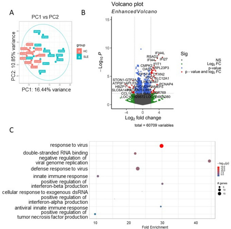
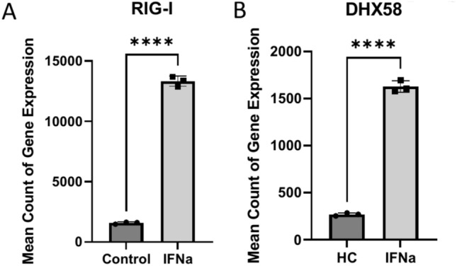
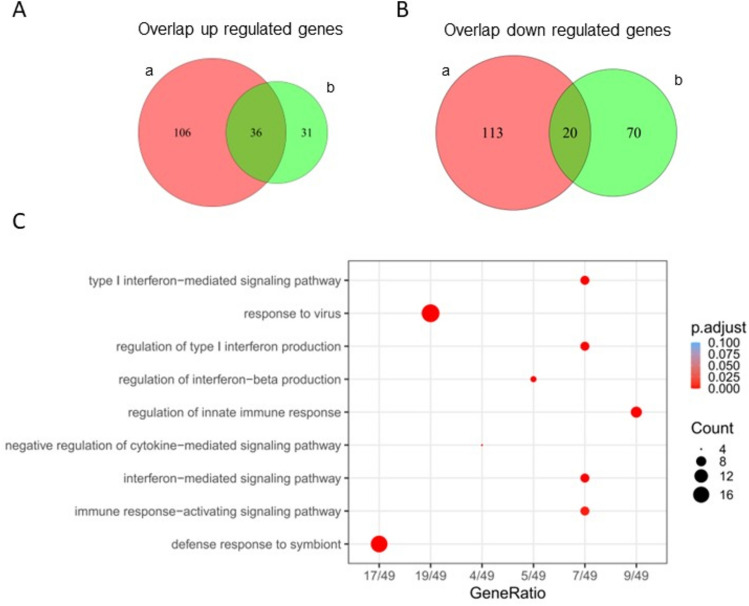
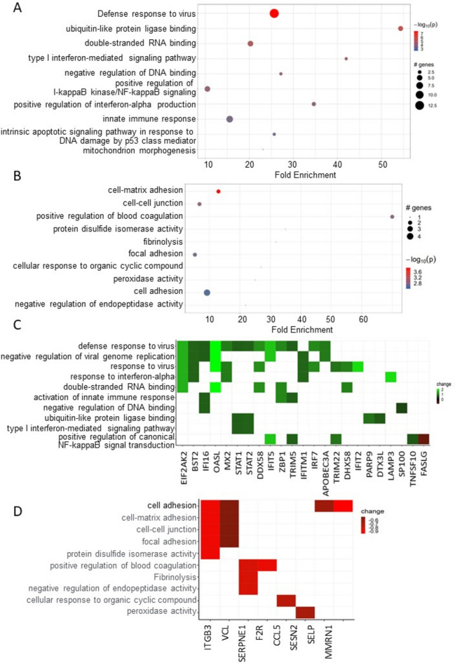
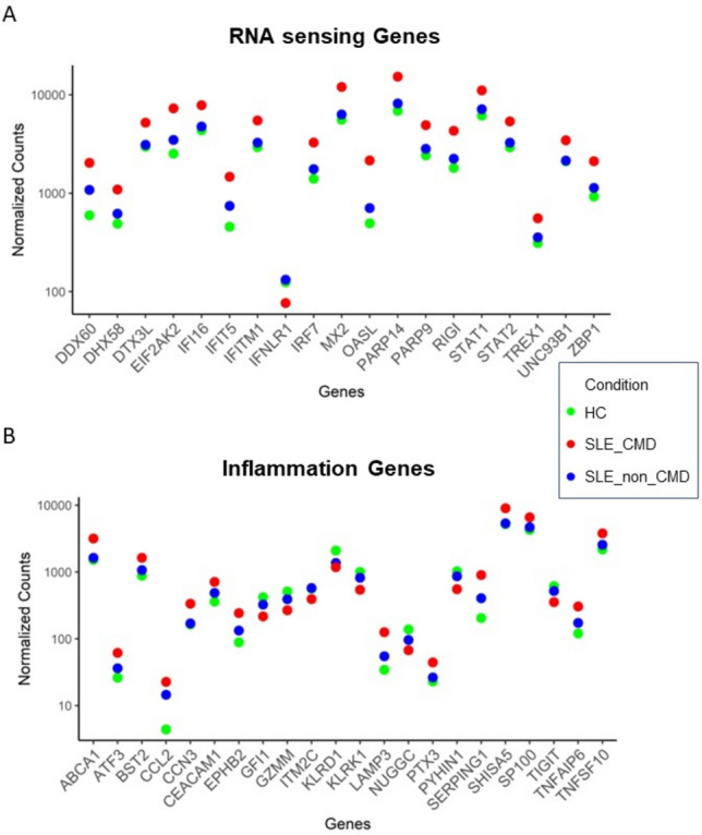
Systemic lupus erythematosus (SLE) patients are 90% women and over three times more likely to die of cardiovascular disease than women in the general population. Chest pain with no obstructive cardiac disease is associated with coronary microvascular disease (CMD), where narrowing of the small blood vessels can lead to ischemia, and frequently reported by SLE patients. Using whole blood RNA samples, we asked whether gene signatures discriminate SLE patients with coronary microvascular dysfunction (CMD) on cardiac MRI (n = 4) from those without (n = 7) and whether any signaling pathway is linked to the underlying pathobiology of SLE CMD. RNA-seq analysis revealed 143 differentially expressed (DE) genes between the SLE and healthy control (HC) groups, with virus defense and interferon (IFN) signaling being the key pathways identified as enriched in SLE as expected. We next conducted a comparative analysis of genes differentially expressed in SLE-CMD and SLE-non-CMD relative to HC samples. Our analysis highlighted differences in IFN signaling, RNA sensing and ADP-ribosylation pathways between SLE-CMD and SLE-non-CMD. This is the first study to investigate possible gene signatures associating with CMD in SLE, and our data strongly suggests that distinct molecular mechanisms underly vascular changes in CMD and non-CMD involvement in SLE.
系统性红斑狼疮(SLE)患者中90%为女性,其死于心血管疾病的可能性是普通人群中女性的三倍多。无阻塞性心脏病的胸痛与冠状动脉微血管疾病(CMD)相关,小血管狭窄可导致缺血,SLE患者经常报告此类胸痛。我们使用全血RNA样本,研究基因特征能否区分心脏磁共振成像(MRI)检查显示有冠状动脉微血管功能障碍(CMD)的SLE患者(n = 4)和无此情况的SLE患者(n = 7),以及是否有任何信号通路与SLE CMD的潜在病理生物学相关。RNA测序分析显示,SLE组与健康对照组(HC)之间有143个差异表达基因,正如预期的那样,病毒防御和干扰素(IFN)信号通路是在SLE中富集的关键通路。接下来,我们对SLE-CMD组和SLE-非CMD组相对于HC样本中差异表达的基因进行了比较分析。我们的分析突出了SLE-CMD组和SLE-非CMD组在IFN信号通路、RNA传感和ADP-核糖基化途径方面的差异。这是第一项研究SLE中与CMD相关的可能基因特征的研究,我们的数据有力地表明,CMD和非CMD累及SLE时血管变化的潜在分子机制不同。