Foti Roberta, Zeppieri Marco, Foti Rosario, Bosco Ylenia Dal, Scollo Davide, Visalli Elisa, Ficili Salvatore, Amato Giorgio, Cifalinò Valentina, Foti Riccardo, Avitabile Alessandro, Cannizzaro Ludovica, Gagliano Caterina
Division of Rheumatology, Policlinico-San Marco Academic Hospital (AOU), Catania 95123, Italy.
Department of Ophthalmology, University Hospital of Udine, Udine 33100, Italy.
J Biol Methods. 2024 Nov 7;11(4):e99010034. doi: 10.14440/jbm.2024.0049. eCollection 2024.
Anterior uveitis is a common manifestation in individuals with rheumatic conditions such as spondylarthritis, Behçet's syndrome, juvenile idiopathic arthritis, and sarcoidosis. Clinical differentiation between granulomatous and non-granulomatous corneal endothelial exudates is crucial to subsequent diagnosis and treatment. Anterior segment optical coherence tomography (AS-OCT) can ensure an accurate differential diagnosis and appropriate follow-up after local and systemic therapy.
This study aimed to distinguish between granulomatous and non-granulomatous endothelial exudates in patients with anterior uveitis using AS-OCT.
This longitudinal observational study involved 30 patients diagnosed with or suspected of having rheumatic autoimmune disease presenting with anterior uveitis. The study was conducted at the combined Rheumatology and Ophthalmology Clinic, San Marco Hospital, Catania, Italy. All patients underwent slit-lamp examination, which revealed or suspected corneal endothelial exudates. A comprehensive rheumatological and ophthalmological evaluation was also performed. Subsequently, the patients were subjected to AS-OCT using the Optovue Solix device.
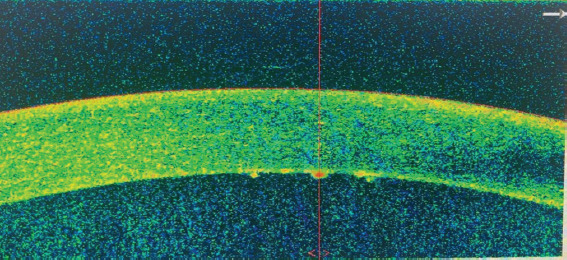
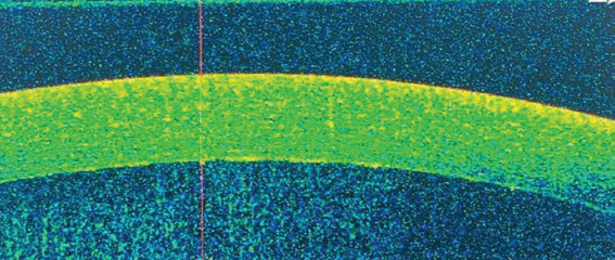
Granulomatous corneal exudates were identified in 30% of the subjects, with counts ranging from 5 to 20 and sizes varying between 50 and 150 μm. Detailed 3D scans further exhibited the morphology of these exudates. A follow-up of patients after steroid therapy (both topical and systemic) and immunosuppressive treatment demonstrated a progressive reduction in the exudates, ultimately leading to their complete resolution.
Use of ophthalmological equipment that allows for simple, rapid, and non-invasive investigations in combination with a multidisciplinary approach, enables appropriate diagnosis and monitoring of therapeutic efficacy in patients with inflammatory ocular conditions presenting with corneal endothelial exudates.
前葡萄膜炎是脊柱关节炎、白塞病、幼年特发性关节炎和结节病等风湿性疾病患者的常见表现。肉芽肿性和非肉芽肿性角膜内皮渗出物的临床鉴别对于后续诊断和治疗至关重要。眼前节光学相干断层扫描(AS-OCT)可确保在局部和全身治疗后进行准确的鉴别诊断和适当的随访。
本研究旨在使用AS-OCT区分前葡萄膜炎患者的肉芽肿性和非肉芽肿性内皮渗出物。
这项纵向观察性研究纳入了30例诊断为或疑似患有风湿性自身免疫性疾病并伴有前葡萄膜炎的患者。该研究在意大利卡塔尼亚圣马尔科医院的风湿科和眼科联合门诊进行。所有患者均接受裂隙灯检查,发现或疑似有角膜内皮渗出物。还进行了全面的风湿科和眼科评估。随后,使用Optovue Solix设备对患者进行AS-OCT检查。
30%的受试者中发现肉芽肿性角膜渗出物,数量在5至20个之间,大小在50至150μm之间。详细的三维扫描进一步显示了这些渗出物的形态。对患者进行局部和全身类固醇治疗以及免疫抑制治疗后的随访显示,渗出物逐渐减少,最终完全消退。
使用能够进行简单、快速且无创检查的眼科设备,并结合多学科方法,能够对出现角膜内皮渗出物的炎症性眼病患者进行适当的诊断和治疗效果监测。