• 文献检索
  • 文档翻译
  • 深度研究
  • 学术资讯
  • Suppr Zotero 插件Zotero 插件
  • 邀请有礼
  • 套餐&价格
  • 历史记录
应用&插件
Suppr Zotero 插件Zotero 插件浏览器插件Mac 客户端Windows 客户端微信小程序
定价
高级版会员购买积分包购买API积分包
服务
文献检索文档翻译深度研究API 文档MCP 服务
关于我们
关于 Suppr公司介绍联系我们用户协议隐私条款
关注我们

Suppr 超能文献

核心技术专利:CN118964589B侵权必究
粤ICP备2023148730 号-1Suppr @ 2026

文献检索

告别复杂PubMed语法,用中文像聊天一样搜索,搜遍4000万医学文献。AI智能推荐,让科研检索更轻松。

立即免费搜索

文件翻译

保留排版,准确专业,支持PDF/Word/PPT等文件格式,支持 12+语言互译。

免费翻译文档

深度研究

AI帮你快速写综述,25分钟生成高质量综述,智能提取关键信息,辅助科研写作。

立即免费体验

使用moscot对细胞进行时空映射。

Mapping cells through time and space with moscot.

作者信息

Klein Dominik, Palla Giovanni, Lange Marius, Klein Michal, Piran Zoe, Gander Manuel, Meng-Papaxanthos Laetitia, Sterr Michael, Saber Lama, Jing Changying, Bastidas-Ponce Aimée, Cota Perla, Tarquis-Medina Marta, Parikh Shrey, Gold Ilan, Lickert Heiko, Bakhti Mostafa, Nitzan Mor, Cuturi Marco, Theis Fabian J

机构信息

Institute of Computational Biology, Helmholtz Center, Munich, Germany.

Department of Mathematics, Technical University of Munich, Garching, Germany.

出版信息

Nature. 2025 Feb;638(8052):1065-1075. doi: 10.1038/s41586-024-08453-2. Epub 2025 Jan 22.

DOI:10.1038/s41586-024-08453-2
PMID:39843746
原文链接:https://pmc.ncbi.nlm.nih.gov/articles/PMC11864987/
Abstract

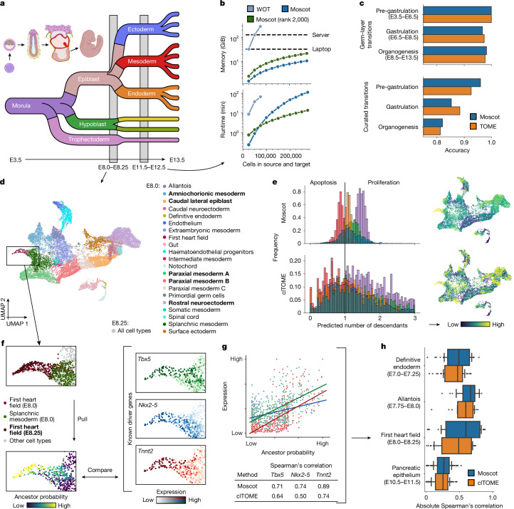
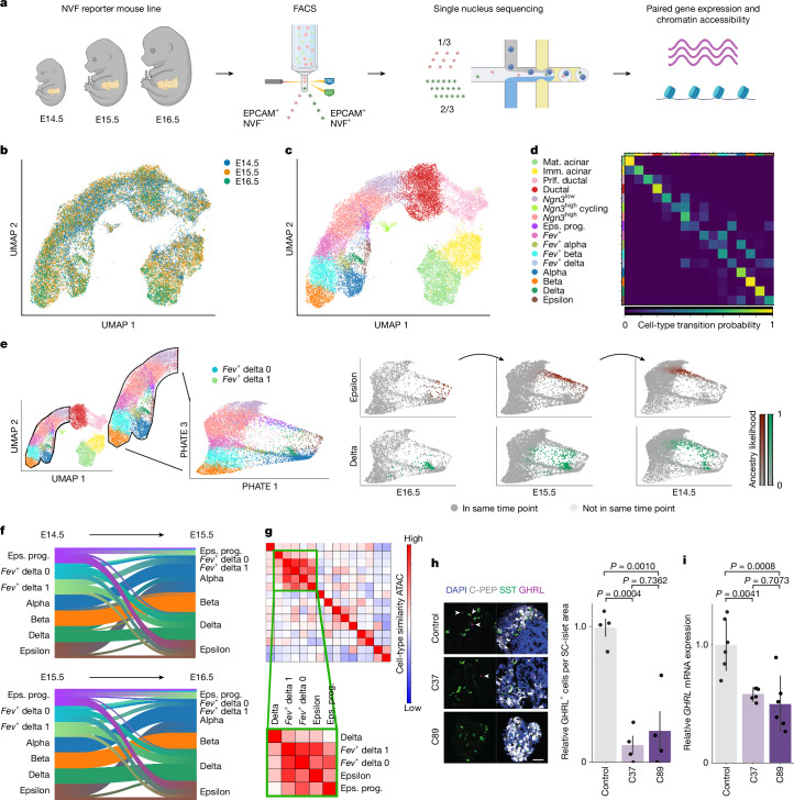
Single-cell genomic technologies enable the multimodal profiling of millions of cells across temporal and spatial dimensions. However, experimental limitations hinder the comprehensive measurement of cells under native temporal dynamics and in their native spatial tissue niche. Optimal transport has emerged as a powerful tool to address these constraints and has facilitated the recovery of the original cellular context. Yet, most optimal transport applications are unable to incorporate multimodal information or scale to single-cell atlases. Here we introduce multi-omics single-cell optimal transport (moscot), a scalable framework for optimal transport in single-cell genomics that supports multimodality across all applications. We demonstrate the capability of moscot to efficiently reconstruct developmental trajectories of 1.7 million cells from mouse embryos across 20 time points. To illustrate the capability of moscot in space, we enrich spatial transcriptomic datasets by mapping multimodal information from single-cell profiles in a mouse liver sample and align multiple coronal sections of the mouse brain. We present moscot.spatiotemporal, an approach that leverages gene-expression data across both spatial and temporal dimensions to uncover the spatiotemporal dynamics of mouse embryogenesis. We also resolve endocrine-lineage relationships of delta and epsilon cells in a previously unpublished mouse, time-resolved pancreas development dataset using paired measurements of gene expression and chromatin accessibility. Our findings are confirmed through experimental validation of NEUROD2 as a regulator of epsilon progenitor cells in a model of human induced pluripotent stem cell islet cell differentiation. Moscot is available as open-source software, accompanied by extensive documentation.

摘要

单细胞基因组技术能够在时间和空间维度上对数百万个细胞进行多模态分析。然而,实验限制阻碍了对处于自然时间动态及其自然空间组织微环境中的细胞进行全面测量。最优传输已成为解决这些限制的有力工具,并有助于恢复原始细胞环境。然而,大多数最优传输应用无法整合多模态信息或扩展到单细胞图谱。在这里,我们介绍了多组学单细胞最优传输(moscot),这是一种用于单细胞基因组学中最优传输的可扩展框架,支持所有应用中的多模态。我们展示了moscot能够有效地重建来自小鼠胚胎的170万个细胞在20个时间点的发育轨迹。为了说明moscot在空间方面的能力,我们通过映射小鼠肝脏样本中单细胞图谱的多模态信息来丰富空间转录组数据集,并对齐小鼠大脑的多个冠状切片。我们提出了moscot.spatiotemporal,一种利用跨空间和时间维度的基因表达数据来揭示小鼠胚胎发生时空动态的方法。我们还使用基因表达和染色质可及性的配对测量,解析了一个此前未发表的小鼠时间分辨胰腺发育数据集中δ细胞和ε细胞的内分泌谱系关系。我们的发现通过在人类诱导多能干细胞胰岛细胞分化模型中对NEUROD2作为ε祖细胞调节剂的实验验证得到了证实。moscot作为开源软件提供,并附有详细文档。

https://cdn.ncbi.nlm.nih.gov/pmc/blobs/52d8/11864987/9aba570cd225/41586_2024_8453_Fig15_ESM.jpg
https://cdn.ncbi.nlm.nih.gov/pmc/blobs/52d8/11864987/83b1429fdb22/41586_2024_8453_Fig1_HTML.jpg
https://cdn.ncbi.nlm.nih.gov/pmc/blobs/52d8/11864987/97f1f7b4e4e1/41586_2024_8453_Fig2_HTML.jpg
https://cdn.ncbi.nlm.nih.gov/pmc/blobs/52d8/11864987/ea67525aef5e/41586_2024_8453_Fig3_HTML.jpg
https://cdn.ncbi.nlm.nih.gov/pmc/blobs/52d8/11864987/6f0a3aa8eb5b/41586_2024_8453_Fig4_HTML.jpg
https://cdn.ncbi.nlm.nih.gov/pmc/blobs/52d8/11864987/792a996474a7/41586_2024_8453_Fig5_HTML.jpg
https://cdn.ncbi.nlm.nih.gov/pmc/blobs/52d8/11864987/5c6af13617e0/41586_2024_8453_Fig6_ESM.jpg
https://cdn.ncbi.nlm.nih.gov/pmc/blobs/52d8/11864987/a2975291fb18/41586_2024_8453_Fig7_ESM.jpg
https://cdn.ncbi.nlm.nih.gov/pmc/blobs/52d8/11864987/2a418d0cd2af/41586_2024_8453_Fig8_ESM.jpg
https://cdn.ncbi.nlm.nih.gov/pmc/blobs/52d8/11864987/8e7f0711edde/41586_2024_8453_Fig9_ESM.jpg
https://cdn.ncbi.nlm.nih.gov/pmc/blobs/52d8/11864987/ce0854ef71e5/41586_2024_8453_Fig10_ESM.jpg
https://cdn.ncbi.nlm.nih.gov/pmc/blobs/52d8/11864987/db78a6d33fb9/41586_2024_8453_Fig11_ESM.jpg
https://cdn.ncbi.nlm.nih.gov/pmc/blobs/52d8/11864987/a2c11c12ebb1/41586_2024_8453_Fig12_ESM.jpg
https://cdn.ncbi.nlm.nih.gov/pmc/blobs/52d8/11864987/7ce858651350/41586_2024_8453_Fig13_ESM.jpg
https://cdn.ncbi.nlm.nih.gov/pmc/blobs/52d8/11864987/489eba9216c5/41586_2024_8453_Fig14_ESM.jpg
https://cdn.ncbi.nlm.nih.gov/pmc/blobs/52d8/11864987/9aba570cd225/41586_2024_8453_Fig15_ESM.jpg

相似文献

1
Mapping cells through time and space with moscot.使用moscot对细胞进行时空映射。
Nature. 2025 Feb;638(8052):1065-1075. doi: 10.1038/s41586-024-08453-2. Epub 2025 Jan 22.
2
Multiplexed spatial mapping of chromatin features, transcriptome and proteins in tissues.组织中染色质特征、转录组和蛋白质的多重空间图谱分析。
Nat Methods. 2025 Mar;22(3):520-529. doi: 10.1038/s41592-024-02576-0. Epub 2025 Jan 27.
3
Retrospective analysis: reproducibility of interblastomere differences of mRNA expression in 2-cell stage mouse embryos is remarkably poor due to combinatorial mechanisms of blastomere diversification.回顾性分析:由于囊胚细胞多样化的组合机制,2 细胞期小鼠胚胎中 mRNA 表达的卵裂球间差异的可重复性极差。
Mol Hum Reprod. 2018 Jul 1;24(7):388-400. doi: 10.1093/molehr/gay021.
4
Cellular diversity and lineage trajectory: insights from mouse single cell transcriptomes.细胞多样性和谱系轨迹:来自小鼠单细胞转录组的见解。
Development. 2020 Jan 24;147(2):dev179788. doi: 10.1242/dev.179788.
5
Next-Generation Lineage Tracing and Fate Mapping to Interrogate Development.下一代谱系追踪和命运图谱解析发育。
Dev Cell. 2021 Jan 11;56(1):7-21. doi: 10.1016/j.devcel.2020.10.021. Epub 2020 Nov 19.
6
Embryo-scale, single-cell spatial transcriptomics.胚胎尺度单细胞空间转录组学
Science. 2021 Jul 2;373(6550):111-117. doi: 10.1126/science.abb9536.
7
Quantitative lineage analysis identifies a hepato-pancreato-biliary progenitor niche.定量谱系分析确定了肝胆胰胆管祖细胞龛位。
Nature. 2021 Sep;597(7874):87-91. doi: 10.1038/s41586-021-03844-1. Epub 2021 Aug 25.
8
Deciphering human macrophage development at single-cell resolution.解析单细胞分辨率下人巨噬细胞的发育。
Nature. 2020 Jun;582(7813):571-576. doi: 10.1038/s41586-020-2316-7. Epub 2020 May 20.
9
Identification of genomic enhancers through spatial integration of single-cell transcriptomics and epigenomics.通过单细胞转录组学和表观基因组学的空间整合来鉴定基因组增强子。
Mol Syst Biol. 2020 May;16(5):e9438. doi: 10.15252/msb.20209438.
10
HelPredictor models single-cell transcriptome to predict human embryo lineage allocation.HelPredictor模型通过单细胞转录组预测人类胚胎谱系分配。
Brief Bioinform. 2021 Nov 5;22(6). doi: 10.1093/bib/bbab196.

引用本文的文献

1
Single-cell multiome and spatial profiling reveals pancreas cell type-specific gene regulatory programs of type 1 diabetes progression.单细胞多组学和空间分析揭示了1型糖尿病进展中胰腺细胞类型特异性基因调控程序。
Sci Adv. 2025 Sep 12;11(37):eady0080. doi: 10.1126/sciadv.ady0080. Epub 2025 Sep 10.
2
Quantifying Landscape and Flux from Single-Cell Omics: Unraveling the Physical Mechanisms of Cell Function.量化单细胞组学中的景观与通量:揭示细胞功能的物理机制
JACS Au. 2025 Aug 7;5(8):3738-3757. doi: 10.1021/jacsau.5c00620. eCollection 2025 Aug 25.
3
Fluctuation structure predicts genome-wide perturbation outcomes.

本文引用的文献

1
Insulin regulates human pancreatic endocrine cell differentiation in vitro.胰岛素在体外调节人胰腺内分泌细胞分化。
Mol Metab. 2024 Jan;79:101853. doi: 10.1016/j.molmet.2023.101853. Epub 2023 Dec 14.
2
Causal identification of single-cell experimental perturbation effects with CINEMA-OT.利用 CINEMA-OT 进行单细胞实验扰动影响的因果识别。
Nat Methods. 2023 Nov;20(11):1769-1779. doi: 10.1038/s41592-023-02040-5. Epub 2023 Nov 2.
3
Deep generative modeling of transcriptional dynamics for RNA velocity analysis in single cells.
波动结构可预测全基因组扰动结果。
Res Sq. 2025 Aug 12:rs.3.rs-7304871. doi: 10.21203/rs.3.rs-7304871/v1.
4
Winnow-KAN: single-cell RNA-seq location recovery with small-gene-set spatial transcriptomics.Winnow-KAN:利用小基因集空间转录组学进行单细胞RNA测序定位恢复
BMC Bioinformatics. 2025 Aug 12;26(1):209. doi: 10.1186/s12859-025-06243-9.
5
Deciphering spatially confined immune evasion niches in osteosarcoma with 3-D spatial transcriptomics: a literature review.利用三维空间转录组学解析骨肉瘤中空间受限的免疫逃逸微环境:文献综述
Front Oncol. 2025 Jul 16;15:1640645. doi: 10.3389/fonc.2025.1640645. eCollection 2025.
6
A variational deep-learning approach to modeling memory T cell dynamics.一种用于模拟记忆性T细胞动力学的变分深度学习方法。
PLoS Comput Biol. 2025 Jul 24;21(7):e1013242. doi: 10.1371/journal.pcbi.1013242.
7
Datavzrd: Rapid programming- and maintenance-free interactive visualization and communication of tabular data.Datavzrd:无需编程和维护即可快速实现表格数据的交互式可视化与通信。
PLoS One. 2025 Jul 22;20(7):e0323079. doi: 10.1371/journal.pone.0323079. eCollection 2025.
8
The tumor microenvironment across four dimensions: assessing space and time in cancer biology.肿瘤微环境的四个维度:评估癌症生物学中的空间与时间
Front Immunol. 2025 Jun 23;16:1554114. doi: 10.3389/fimmu.2025.1554114. eCollection 2025.
9
Computational modeling of single-cell dynamics data.单细胞动力学数据的计算建模。
Brief Bioinform. 2025 May 1;26(3). doi: 10.1093/bib/bbaf305.
10
Evaluating discrepancies in dimensionality reduction for time-series single-cell RNA-sequencing data.评估时间序列单细胞RNA测序数据降维中的差异。
Brief Bioinform. 2025 May 1;26(3). doi: 10.1093/bib/bbaf287.
单细胞 RNA 速度分析的转录动态深度生成模型。
Nat Methods. 2024 Jan;21(1):50-59. doi: 10.1038/s41592-023-01994-w. Epub 2023 Sep 21.
4
The specious art of single-cell genomics.单细胞基因组学的似是而非的艺术。
PLoS Comput Biol. 2023 Aug 17;19(8):e1011288. doi: 10.1371/journal.pcbi.1011288. eCollection 2023 Aug.
5
Partial alignment of multislice spatially resolved transcriptomics data.多切片空间分辨转录组学数据的部分比对。
Genome Res. 2023 Jul;33(7):1124-1132. doi: 10.1101/gr.277670.123. Epub 2023 Aug 8.
6
SCENIC+: single-cell multiomic inference of enhancers and gene regulatory networks.SCENIC+:单细胞多组学推断增强子和基因调控网络。
Nat Methods. 2023 Sep;20(9):1355-1367. doi: 10.1038/s41592-023-01938-4. Epub 2023 Jul 13.
7
Multi-omic single-cell velocity models epigenome-transcriptome interactions and improves cell fate prediction.多组学单细胞速度模型揭示了表观基因组-转录组相互作用,并提高了细胞命运预测。
Nat Biotechnol. 2023 Mar;41(3):387-398. doi: 10.1038/s41587-022-01476-y. Epub 2022 Oct 13.
8
Single-cell Stereo-seq reveals induced progenitor cells involved in axolotl brain regeneration.单细胞立体测序揭示参与蝾螈大脑再生的诱导祖细胞。
Science. 2022 Sep 2;377(6610):eabp9444. doi: 10.1126/science.abp9444.
9
Spatial profiling of chromatin accessibility in mouse and human tissues.在小鼠和人体组织中进行染色质可及性的空间分析。
Nature. 2022 Sep;609(7926):375-383. doi: 10.1038/s41586-022-05094-1. Epub 2022 Aug 17.
10
Towards a comprehensive evaluation of dimension reduction methods for transcriptomic data visualization.面向转录组数据可视化的降维方法综合评估。
Commun Biol. 2022 Jul 19;5(1):719. doi: 10.1038/s42003-022-03628-x.