Saroj Smrithi, Vijayalakshmi U
Department of Chemistry, School of Advanced Sciences, Vellore Institute of Technology, Vellore, Tamil Nadu, 632014, India.
Sci Rep. 2025 Jan 25;15(1):3234. doi: 10.1038/s41598-025-87111-7.
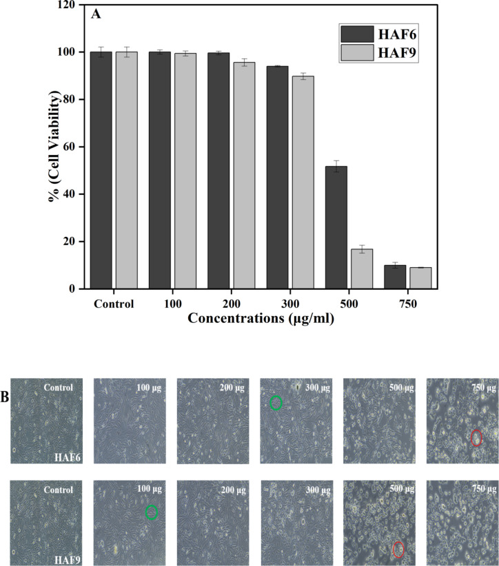
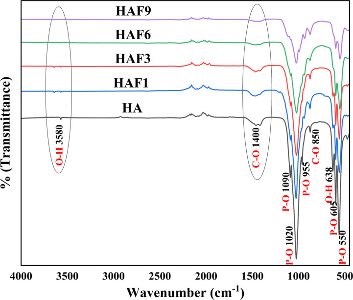
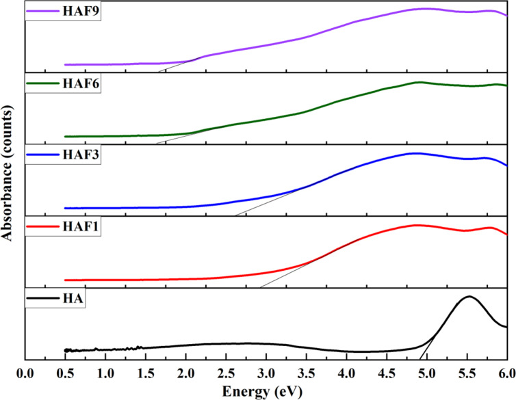
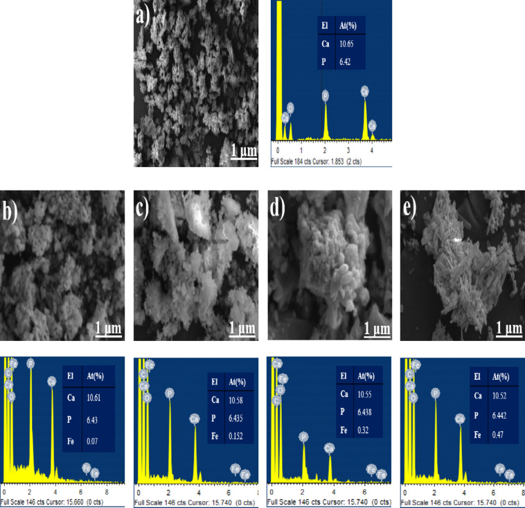
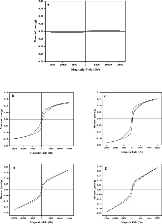
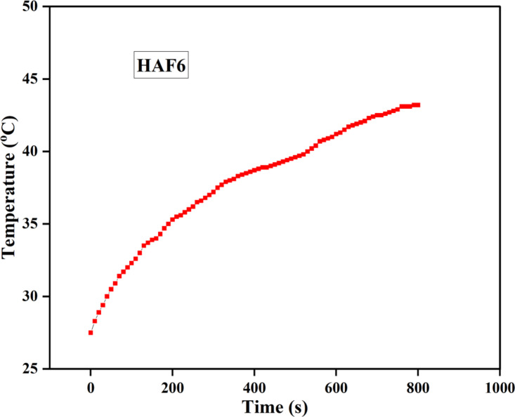
Hydroxyapatite (HA) is an important constituent of natural bone. The properties of HA can be enhanced with the help of various ionic substitutions in the crystal lattice of HA. Iron (Fe) is a vital element present in bones and teeth. In this study, iron-doped HA was synthesized using a refluxing-based sol-gel route with varying concentrations of iron (1-9 M%). Samples were analyzed using an X-ray diffractometer (XRD), UV-Vis Spectrophotometer, Fourier-transform infrared spectroscopy (FT-IR), vibrating sample magnetometer (VSM) and Scanning Electron Microscope (SEM). The biological assessment was carried out by hemolytic assay, anti-bacterial activity and in-vitro biocompatibility. XRD data confirmed the evolution of the hexagonal HA crystal structure with the reduction in the crystallinity and the crystallite size. All the characteristic bands were confirmed using FT-IR which also further proved the existence of A-type carbonated apatite. The UV-Vis spectra confirmed the reduction in the band gap energies owing to the substitution of iron. The SEM results showed a change in the shape of the samples with increasing iron concentration. The magnetic behavior of samples also altered from diamagnetic to ferromagnetic behavior due to the doping of iron with enhanced heating efficiency. All the samples were found to be hemocompatible. The antibacterial efficacy was found to be higher for E. coli (gram-negative) bacteria compared to S. aureus (gram-positive) bacteria. Moreover, the superior cell viability of MG-63 (osteoblast-like) cells was observed in Fe-doped HA, attributed to MTT assay which revealed the enhanced cell viability of osteoblast-like cells in the Fe-doped HA. These results strongly emphasize the potential of the developed samples for bone regeneration applications.
羟基磷灰石(HA)是天然骨的重要组成部分。通过在HA晶格中进行各种离子取代,可以增强HA的性能。铁(Fe)是骨骼和牙齿中存在的重要元素。在本研究中,采用基于回流的溶胶-凝胶法合成了不同铁浓度(1-9 M%)的铁掺杂HA。使用X射线衍射仪(XRD)、紫外-可见分光光度计、傅里叶变换红外光谱仪(FT-IR)、振动样品磁强计(VSM)和扫描电子显微镜(SEM)对样品进行了分析。通过溶血试验、抗菌活性和体外生物相容性进行生物学评估。XRD数据证实了六方HA晶体结构的演变,同时结晶度和晶粒尺寸降低。FT-IR证实了所有特征峰,这也进一步证明了A型碳酸化磷灰石的存在。紫外-可见光谱证实了由于铁的取代,带隙能量降低。SEM结果表明,随着铁浓度的增加,样品形状发生了变化。由于铁的掺杂提高了加热效率,样品的磁行为也从抗磁性转变为铁磁性。所有样品均具有血液相容性。发现对大肠杆菌(革兰氏阴性菌)的抗菌效果比对金黄色葡萄球菌(革兰氏阳性菌)更高。此外,在铁掺杂HA中观察到MG-63(成骨样)细胞具有优异的细胞活力,MTT试验表明铁掺杂HA中成骨样细胞的细胞活力增强。这些结果强烈强调了所开发样品在骨再生应用中的潜力。