Haberl David, Ning Jing, Kluge Kilian, Kumpf Katarina, Yu Josef, Jiang Zewen, Constantino Claudia, Monaci Alice, Starace Maria, Haug Alexander R, Calabretta Raffaella, Camoni Luca, Bertagna Francesco, Mascherbauer Katharina, Hofer Felix, Albano Domenico, Sciagra Roberto, Oliveira Francisco, Costa Durval, Nitsche Christian, Hacker Marcus, Spielvogel Clemens P
Department of Biomedical Imaging and Image-guided Therapy, Division of Nuclear Medicine, Medical University of Vienna, Spitalgasse 23, Vienna, 1090, Austria.
Christian Doppler Laboratory for Applied Metabolomics, Medical University of Vienna, Vienna, Austria.
Eur J Nucl Med Mol Imaging. 2025 Jun;52(7):2355-2368. doi: 10.1007/s00259-025-07091-8. Epub 2025 Jan 29.
Advancements of deep learning in medical imaging are often constrained by the limited availability of large, annotated datasets, resulting in underperforming models when deployed under real-world conditions. This study investigated a generative artificial intelligence (AI) approach to create synthetic medical images taking the example of bone scintigraphy scans, to increase the data diversity of small-scale datasets for more effective model training and improved generalization.
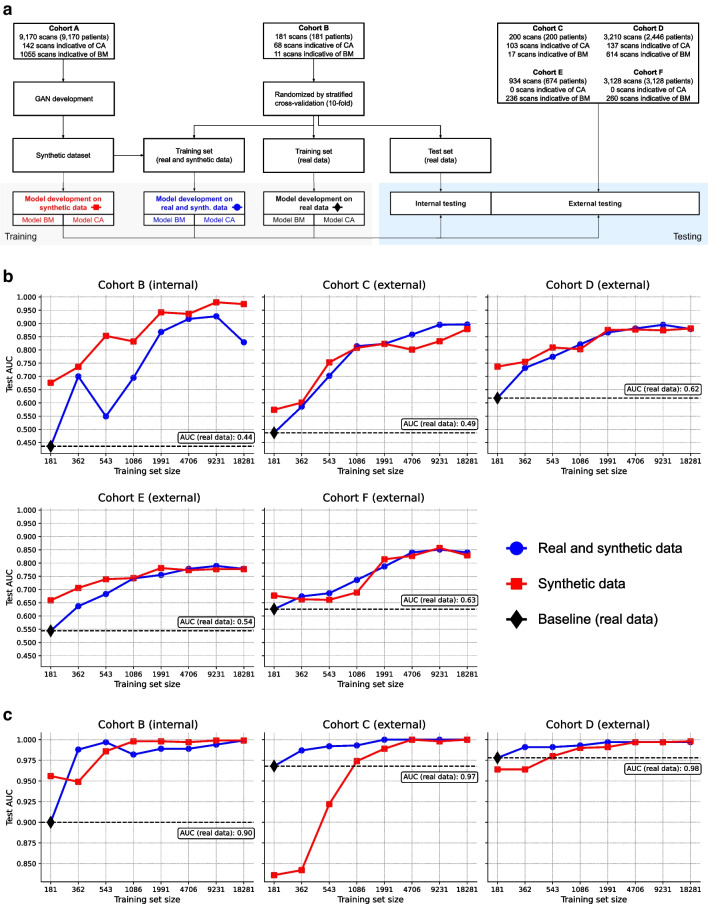
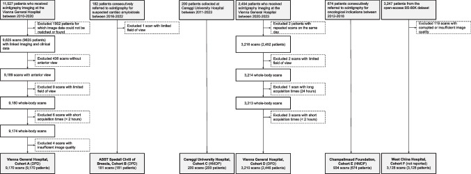
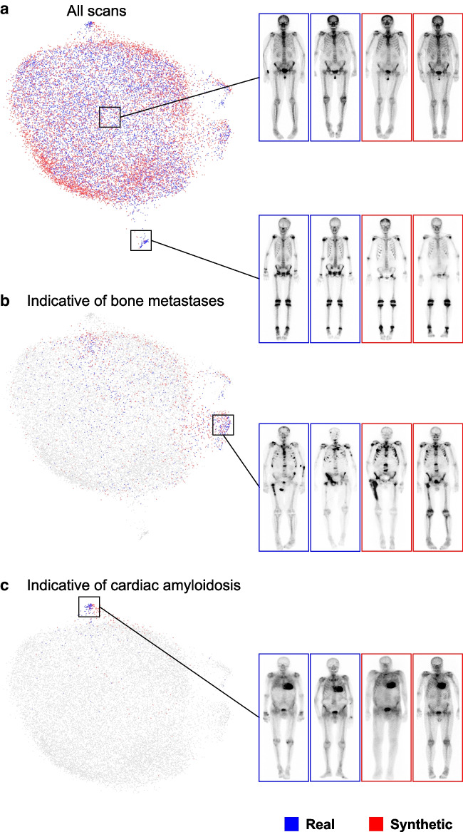
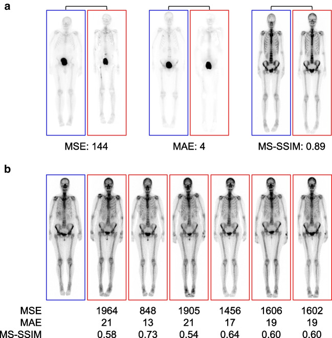
We trained a generative model on Tc-bone scintigraphy scans from 9,170 patients in one center to generate high-quality and fully anonymized annotated scans of patients representing two distinct disease patterns: abnormal uptake indicative of (i) bone metastases and (ii) cardiac uptake indicative of cardiac amyloidosis. A blinded reader study was performed to assess the clinical validity and quality of the generated data. We investigated the added value of the generated data by augmenting an independent small single-center dataset with synthetic data and by training a deep learning model to detect abnormal uptake in a downstream classification task. We tested this model on 7,472 scans from 6,448 patients across four external sites in a cross-tracer and cross-scanner setting and associated the resulting model predictions with clinical outcomes.
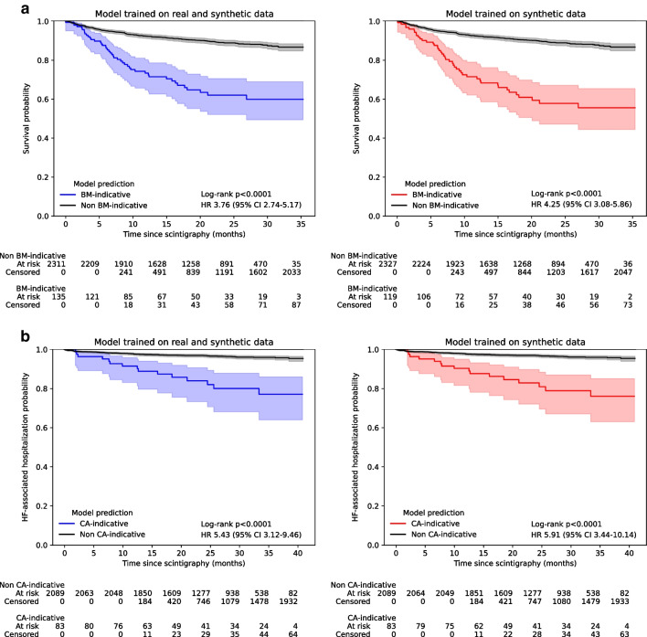
The clinical value and high quality of the synthetic imaging data were confirmed by four readers, who were unable to distinguish synthetic scans from real scans (average accuracy: 0.48% [95% CI 0.46-0.51]), disagreeing in 239 (60%) of 400 cases (Fleiss' kappa: 0.18). Adding synthetic data to the training set improved model performance by a mean (± SD) of 33(± 10)% AUC (p < 0.0001) for detecting abnormal uptake indicative of bone metastases and by 5(± 4)% AUC (p < 0.0001) for detecting uptake indicative of cardiac amyloidosis across both internal and external testing cohorts, compared to models without synthetic training data. Patients with predicted abnormal uptake had adverse clinical outcomes (log-rank: p < 0.0001).
Generative AI enables the targeted generation of bone scintigraphy images representing different clinical conditions. Our findings point to the potential of synthetic data to overcome challenges in data sharing and in developing reliable and prognostic deep learning models in data-limited environments.
深度学习在医学成像领域的进展常常受到大型标注数据集有限可用性的限制,导致在实际应用中模型表现不佳。本研究以骨闪烁扫描为例,探讨了一种生成式人工智能(AI)方法来创建合成医学图像,以增加小规模数据集的数据多样性,从而实现更有效的模型训练和更好的泛化能力。
我们在一个中心的9170例患者的Tc骨闪烁扫描数据上训练了一个生成模型,以生成高质量且完全匿名的标注扫描图像,这些图像代表两种不同的疾病模式:(i)提示骨转移的异常摄取和(ii)提示心脏淀粉样变性的心脏摄取。进行了一项盲法阅片者研究,以评估生成数据的临床有效性和质量。我们通过用合成数据扩充一个独立的小型单中心数据集,并在下游分类任务中训练一个深度学习模型来检测异常摄取,研究了生成数据的附加价值。我们在跨示踪剂和跨扫描仪设置下,对来自四个外部站点的6448例患者的7472次扫描进行了测试,并将所得模型预测结果与临床结果相关联。
四位阅片者证实了合成成像数据的临床价值和高质量,他们无法区分合成扫描图像和真实扫描图像(平均准确率:0.48% [95% CI 0.46 - 0.51]),在400例病例中有239例(60%)存在分歧(Fleiss' kappa:0.18)。与没有合成训练数据的模型相比,在内部和外部测试队列中,将合成数据添加到训练集中可使检测提示骨转移的异常摄取的模型性能平均(±标准差)提高33(±10)%的AUC(p < 0.0001),检测提示心脏淀粉样变性的摄取的模型性能提高5(±4)%的AUC(p < 0.0001)。预测有异常摄取的患者具有不良临床结局(对数秩检验:p < 0.0001)。
生成式AI能够针对性地生成代表不同临床情况的骨闪烁扫描图像。我们的研究结果表明,合成数据在克服数据共享挑战以及在数据有限的环境中开发可靠的预后深度学习模型方面具有潜力。