• 文献检索
  • 文档翻译
  • 深度研究
  • 学术资讯
  • Suppr Zotero 插件Zotero 插件
  • 邀请有礼
  • 套餐&价格
  • 历史记录
应用&插件
Suppr Zotero 插件Zotero 插件浏览器插件Mac 客户端Windows 客户端微信小程序
定价
高级版会员购买积分包购买API积分包
服务
文献检索文档翻译深度研究API 文档MCP 服务
关于我们
关于 Suppr公司介绍联系我们用户协议隐私条款
关注我们

Suppr 超能文献

核心技术专利:CN118964589B侵权必究
粤ICP备2023148730 号-1Suppr @ 2026

文献检索

告别复杂PubMed语法,用中文像聊天一样搜索,搜遍4000万医学文献。AI智能推荐,让科研检索更轻松。

立即免费搜索

文件翻译

保留排版,准确专业,支持PDF/Word/PPT等文件格式,支持 12+语言互译。

免费翻译文档

深度研究

AI帮你快速写综述,25分钟生成高质量综述,智能提取关键信息,辅助科研写作。

立即免费体验

单细胞RNA测序揭示糖尿病足溃疡中表皮分化受损及病理微环境

Single-cell RNA sequencing reveals the impaired epidermal differentiation and pathological microenvironment in diabetic foot ulcer.

作者信息

Liu Yiling, Wang Peng, Li Jingting, Chen Lei, Shu Bin, Wang Hanwen, Liu Hengdeng, Zhao Shixin, Zhou Junli, Chen Xiaodong, Xie Julin

机构信息

Department of Burn and Wound Repair Surgery, The First Affiliated Hospital of Sun Yat-sen University, No. 58 Zhongshan 2 Road, Guangzhou 510080, China.

Institute of Precision Medicine, The First Affiliated Hospital, Sun Yat-Sen University, No. 58, Zhongshan 2 Road, Guangzhou 510080, China.

出版信息

Burns Trauma. 2025 Mar 4;13:tkae065. doi: 10.1093/burnst/tkae065. eCollection 2025.

DOI:10.1093/burnst/tkae065
PMID:40040959
原文链接:https://pmc.ncbi.nlm.nih.gov/articles/PMC11879498/
Abstract

BACKGROUND

Diabetic foot ulcer (DFU) is one of the most common and complex complications of diabetes, but the underlying pathophysiology remains unclear. Single-cell RNA sequencing (scRNA-seq) has been conducted to explore novel cell types or molecular profiles of DFU from various perspectives. This study aimed to comprehensively analyze the potential mechanisms underlying impaired re-epithelization of DFU in a single-cell perspective.

METHODS

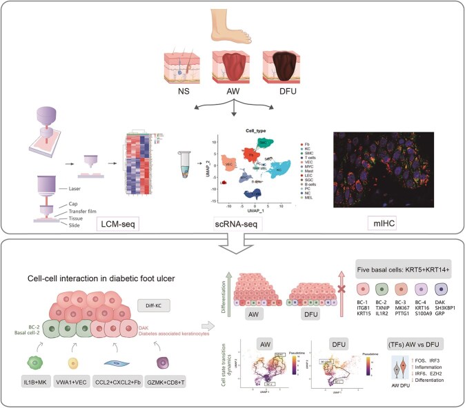
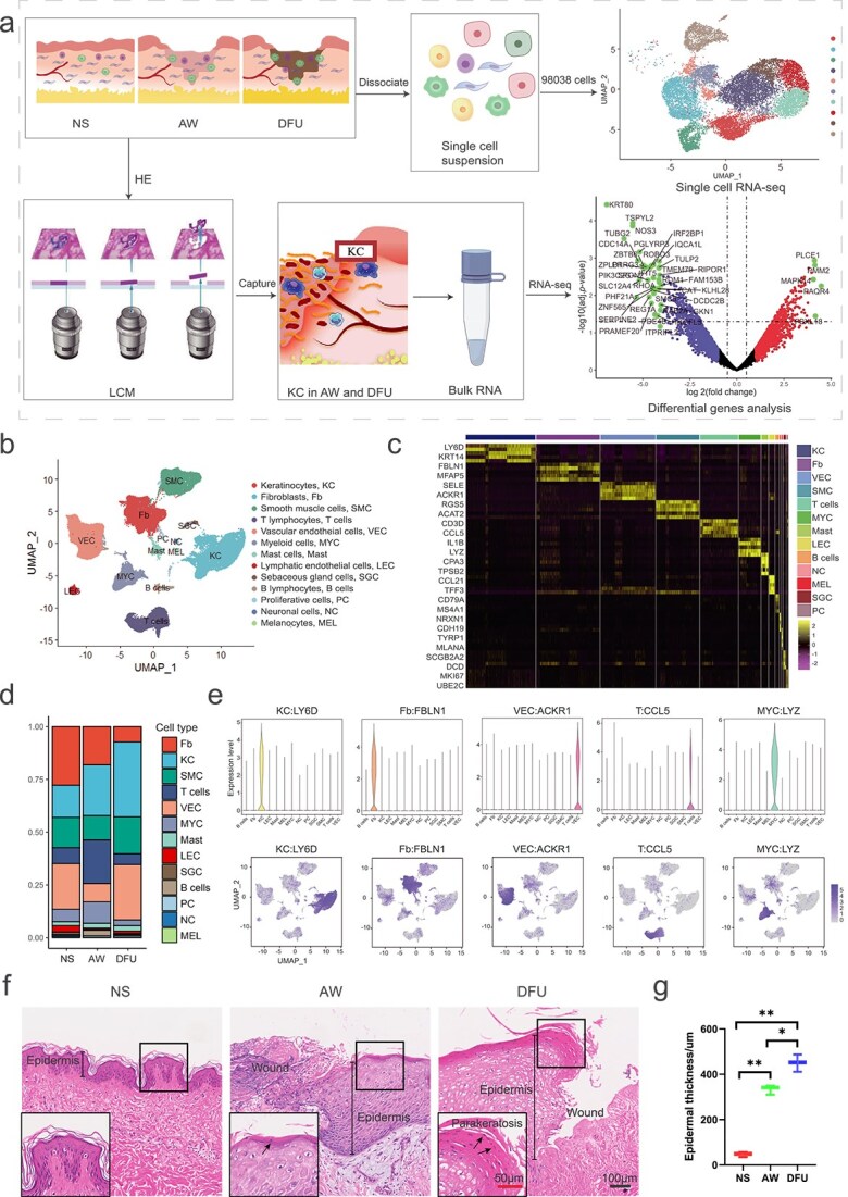
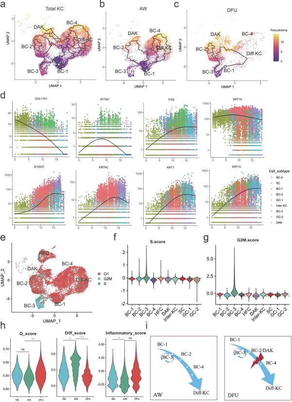
We conducted scRNA-seq on tissues from human normal skin, acute wound, and DFU to investigate the potential mechanisms underlying impaired epidermal differentiation and the pathological microenvironment. Pseudo-time and lineage inference analyses revealed the distinct states and transition trajectories of epidermal cells under different conditions. Transcription factor analysis revealed the potential regulatory mechanism of key subtypes of keratinocytes. Cell-cell interaction analysis revealed the regulatory network between the proinflammatory microenvironment and epidermal cells. Laser-capture microscopy coupled with RNA sequencing (LCM-seq) and multiplex immunohistochemistry were used to validate the expression and location of key subtypes of keratinocytes.

RESULTS

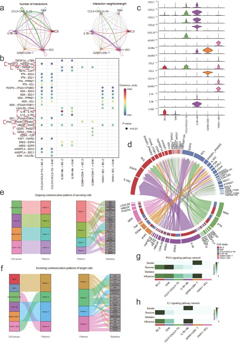
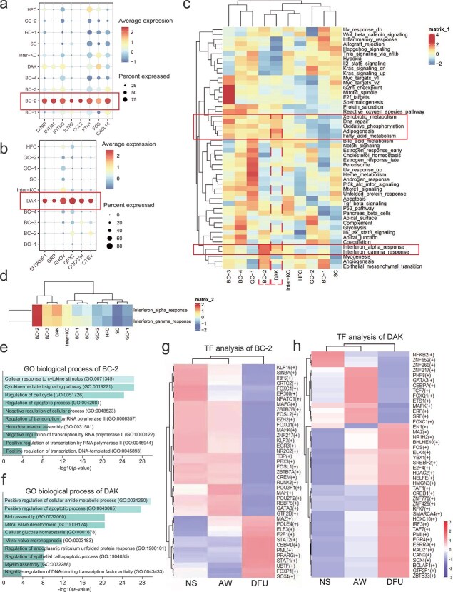
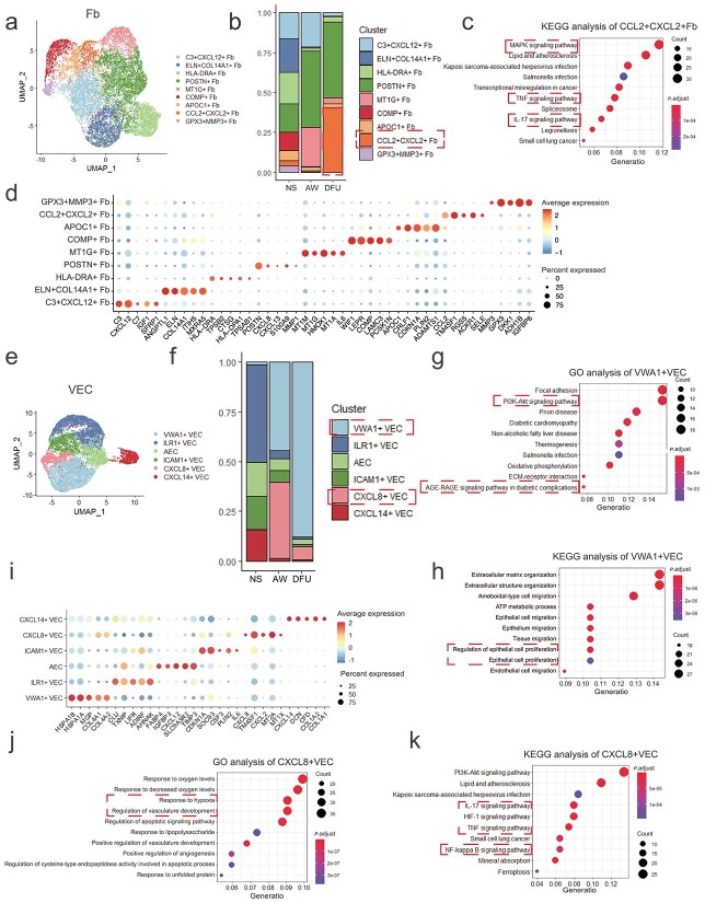
Our research provided a comprehensive map of the phenotypic and dynamic changes that occur during epidermal differentiation, alongside the corresponding regulatory networks in DFU. Importantly, we identified two subtypes of keratinocytes: basal cells (BC-2) and diabetes-associated keratinocytes (DAK) that might play crucial roles in the impairment of epidermal homeostasis. BC-2 and DAK showed a marked increase in DFU, with an inactive state and insufficient motivation for epidermal differentiation. BC-2 was involved in the cellular response and apoptosis processes, with high expression of TXNIP, IFITM1, and IL1R2. Additionally, the pro-differentiation transcription factors were downregulated in BC-2 in DFU, indicating that the differentiation process might be inhibited in BC-2 in DFU. DAK was associated with cellular glucose homeostasis. Furthermore, increased CCL2 + CXCL2+ fibroblasts, VWA1+ vascular endothelial cells, and GZMA+CD8+ T cells were detected in DFU. These changes in the wound microenvironment could regulate the fate of epidermal cells through the TNFSF12-TNFRSF12A, IFNG-IFNGR1/2, and IL-1B-IL1R2 pathways, which might result in persistent inflammation and impaired epidermal differentiation in DFU.

CONCLUSIONS

Our findings offer novel insights into the pathophysiology of DFU and present potential therapeutic targets that could improve wound care and treatment outcomes for DFU patients.

摘要

背景

糖尿病足溃疡(DFU)是糖尿病最常见且复杂的并发症之一,但其潜在的病理生理学机制仍不清楚。已开展单细胞RNA测序(scRNA-seq)从多个角度探索DFU的新型细胞类型或分子特征。本研究旨在从单细胞角度全面分析DFU再上皮化受损的潜在机制。

方法

我们对来自人类正常皮肤、急性伤口和DFU的组织进行scRNA-seq,以研究表皮分化受损和病理微环境的潜在机制。拟时间和谱系推断分析揭示了不同条件下表皮细胞的不同状态和转变轨迹。转录因子分析揭示了角质形成细胞关键亚型的潜在调控机制。细胞-细胞相互作用分析揭示了促炎微环境与表皮细胞之间的调控网络。激光捕获显微镜联合RNA测序(LCM-seq)和多重免疫组化用于验证角质形成细胞关键亚型的表达和定位。

结果

我们的研究提供了一张表皮分化过程中发生的表型和动态变化的综合图谱,以及DFU中相应的调控网络。重要的是,我们鉴定出两种角质形成细胞亚型:基底细胞(BC-2)和糖尿病相关角质形成细胞(DAK),它们可能在表皮稳态受损中起关键作用。BC-2和DAK在DFU中显著增加,处于非活性状态且表皮分化动力不足。BC-2参与细胞反应和凋亡过程,TXNIP、IFITM1和IL1R2表达较高。此外,促分化转录因子在DFU的BC-2中下调,表明DFU中BC-2的分化过程可能受到抑制。DAK与细胞葡萄糖稳态相关。此外,在DFU中检测到CCL2 + CXCL2 +成纤维细胞、VWA1 +血管内皮细胞和GZMA + CD8 + T细胞增加。伤口微环境的这些变化可通过TNFSF12-TNFRSF12A、IFNG-IFNGR1/2和IL-1B-IL1R2途径调节表皮细胞的命运,这可能导致DFU中持续的炎症和表皮分化受损。

结论

我们的研究结果为DFU的病理生理学提供了新的见解,并提出了潜在的治疗靶点,有望改善DFU患者的伤口护理和治疗效果。

https://cdn.ncbi.nlm.nih.gov/pmc/blobs/7549/11879498/8993f410946d/tkae065f6.jpg
https://cdn.ncbi.nlm.nih.gov/pmc/blobs/7549/11879498/9ff7cda15ce0/tkae065ga1.jpg
https://cdn.ncbi.nlm.nih.gov/pmc/blobs/7549/11879498/73fd46ce1151/tkae065f1.jpg
https://cdn.ncbi.nlm.nih.gov/pmc/blobs/7549/11879498/707a8fbbc9fc/tkae065f2.jpg
https://cdn.ncbi.nlm.nih.gov/pmc/blobs/7549/11879498/d1f104057190/tkae065f3.jpg
https://cdn.ncbi.nlm.nih.gov/pmc/blobs/7549/11879498/fdc4560744d3/tkae065f4.jpg
https://cdn.ncbi.nlm.nih.gov/pmc/blobs/7549/11879498/563c82acd335/tkae065f5.jpg
https://cdn.ncbi.nlm.nih.gov/pmc/blobs/7549/11879498/8993f410946d/tkae065f6.jpg

相似文献

1
Single-cell RNA sequencing reveals the impaired epidermal differentiation and pathological microenvironment in diabetic foot ulcer.单细胞RNA测序揭示糖尿病足溃疡中表皮分化受损及病理微环境
Burns Trauma. 2025 Mar 4;13:tkae065. doi: 10.1093/burnst/tkae065. eCollection 2025.
2
Epidermal growth factor effect on lipopolysaccharide-induced inflammation in fibroblasts derived from diabetic foot ulcer.表皮生长因子对糖尿病足溃疡来源的成纤维细胞中脂多糖诱导的炎症的影响
Scars Burn Heal. 2022 Feb 18;8:20595131211067380. doi: 10.1177/20595131211067380. eCollection 2022 Jan-Dec.
3
Single-cell RNA-seq in diabetic foot ulcer wound healing.单细胞 RNA 测序在糖尿病足溃疡愈合中的应用。
Wound Repair Regen. 2024 Nov-Dec;32(6):880-889. doi: 10.1111/wrr.13218. Epub 2024 Sep 12.
4
Single-cell RNA-seq and bulk-seq identify RAB17 as a potential regulator of angiogenesis by human dermal microvascular endothelial cells in diabetic foot ulcers.单细胞RNA测序和批量测序确定RAB17是糖尿病足溃疡中人类真皮微血管内皮细胞血管生成的潜在调节因子。
Burns Trauma. 2023 Aug 18;11:tkad020. doi: 10.1093/burnst/tkad020. eCollection 2023.
5
Sequencing of messenger RNA in the healing process of diabetes foot ulcer.糖尿病足溃疡愈合过程中信使核糖核酸的测序
Front Endocrinol (Lausanne). 2024 Nov 19;15:1468301. doi: 10.3389/fendo.2024.1468301. eCollection 2024.
6
Analysis of immune cell activation in patients with diabetes foot ulcer from the perspective of single cell.从单细胞角度分析糖尿病足溃疡患者的免疫细胞活化情况。
Eur J Med Res. 2024 Dec 19;29(1):606. doi: 10.1186/s40001-024-02179-7.
7
Enhancing diabetic foot ulcer healing: Impact of the regulation of the FUS and ILF2 RNA‑binding proteins through negative pressure wound therapy.增强糖尿病足溃疡愈合:负压伤口疗法对 FUS 和 ILF2 RNA 结合蛋白的调节作用。
Int J Mol Med. 2024 Nov;54(5). doi: 10.3892/ijmm.2024.5427. Epub 2024 Sep 20.
8
Comprehensive transcriptomic analysis of immune-related genes in diabetic foot ulcers: New insights into mechanisms and therapeutic targets.糖尿病足溃疡免疫相关基因的综合转录组学分析:对发病机制和治疗靶点的新认识。
Int Immunopharmacol. 2024 Sep 30;139:112638. doi: 10.1016/j.intimp.2024.112638. Epub 2024 Jul 28.
9
[Screening, functional analysis and clinical validation of differentially expressed genes in diabetic foot ulcers].糖尿病足溃疡中差异表达基因的筛选、功能分析及临床验证
Zhonghua Shao Shang Yu Chuang Mian Xiu Fu Za Zhi. 2022 Oct 20;38(10):944-951. doi: 10.3760/cma.j.cn501225-20220731-00328.
10
[Mechanism of maggot debridement therapy in promoting wound angiogenesis in patients with diabetic foot ulcer].[蛆虫清创疗法促进糖尿病足溃疡患者伤口血管生成的机制]
Zhonghua Shao Shang Za Zhi. 2020 Nov 20;36(11):1040-1049. doi: 10.3760/cma.j.cn501120-20191022-00409.

引用本文的文献

1
Bioinspired wearable polymer microneedle patches: pioneering diabetic wound therapy for the horizon.受生物启发的可穿戴聚合物微针贴片:开创糖尿病伤口治疗的新纪元。
RSC Adv. 2025 Sep 8;15(39):32509-32535. doi: 10.1039/d5ra02557e. eCollection 2025 Sep 5.
2
Single-cell RNA transcriptome analysis reveals regulatory programs of RNA binding proteins in diabetic foot ulcers.单细胞RNA转录组分析揭示糖尿病足溃疡中RNA结合蛋白的调控程序。
Sci Rep. 2025 Aug 23;15(1):31059. doi: 10.1038/s41598-025-16291-z.
3
Exploring machine learning strategies for single-cell transcriptomic analysis in wound healing.

本文引用的文献

1
Analysis of Bulk Transcriptome Sequencing Data and in vitro Experiments Reveal SIN3A as a Potential Target for Diabetic Foot Ulcer.批量转录组测序数据分析与体外实验揭示SIN3A作为糖尿病足溃疡的潜在靶点
Diabetes Metab Syndr Obes. 2023 Dec 18;16:4119-4132. doi: 10.2147/DMSO.S439924. eCollection 2023.
2
Opportunities and tradeoffs in single-cell transcriptomic technologies.单细胞转录组技术中的机遇与权衡
Trends Genet. 2024 Jan;40(1):83-93. doi: 10.1016/j.tig.2023.10.003. Epub 2023 Nov 10.
3
Spatial Transcriptomics: Emerging Technologies in Tissue Gene Expression Profiling.
探索用于伤口愈合单细胞转录组分析的机器学习策略。
Burns Trauma. 2025 May 13;13:tkaf032. doi: 10.1093/burnst/tkaf032. eCollection 2025.
4
Unraveling the oxidative stress landscape in diabetic foot ulcers: insights from bulk RNA and single-cell RNA sequencing data.解析糖尿病足溃疡中的氧化应激状况:来自 bulk RNA 和单细胞 RNA 测序数据的见解
Biol Direct. 2025 Jul 4;20(1):79. doi: 10.1186/s13062-025-00672-5.
5
Immune cell contribution to vascular complications in diabetes.免疫细胞在糖尿病血管并发症中的作用。
Front Endocrinol (Lausanne). 2025 May 21;16:1549945. doi: 10.3389/fendo.2025.1549945. eCollection 2025.
空间转录组学:组织基因表达分析的新兴技术。
Anal Chem. 2023 Oct 24;95(42):15450-15460. doi: 10.1021/acs.analchem.3c02029. Epub 2023 Oct 10.
4
Single-cell RNA-seq and bulk-seq identify RAB17 as a potential regulator of angiogenesis by human dermal microvascular endothelial cells in diabetic foot ulcers.单细胞RNA测序和批量测序确定RAB17是糖尿病足溃疡中人类真皮微血管内皮细胞血管生成的潜在调节因子。
Burns Trauma. 2023 Aug 18;11:tkad020. doi: 10.1093/burnst/tkad020. eCollection 2023.
5
Decoding the Human Epidermal Complexity at Single-Cell Resolution.单细胞分辨率解析人类表皮复杂性。
Int J Mol Sci. 2023 May 10;24(10):8544. doi: 10.3390/ijms24108544.
6
Investigation of the keratinocyte transcriptome altered in high-glucose environment: An in-vitro model system for precision medicine.高糖环境下角质形成细胞转录组改变的研究:一种用于精准医学的体外模型系统。
J Dermatol Sci. 2023 Jan;109(1):37-46. doi: 10.1016/j.jdermsci.2023.01.007. Epub 2023 Jan 18.
7
Macrophage-specific inhibition of the histone demethylase JMJD3 decreases STING and pathologic inflammation in diabetic wound repair.组织特异性抑制组蛋白去甲基化酶 JMJD3 可减少糖尿病伤口修复中的 STING 和病理性炎症。
Cell Mol Immunol. 2022 Nov;19(11):1251-1262. doi: 10.1038/s41423-022-00919-5. Epub 2022 Sep 20.
8
Activation of the STING-IRF3 pathway involved in psoriasis with diabetes mellitus.STING-IRF3 通路的激活与糖尿病合并银屑病有关。
J Cell Mol Med. 2022 Apr;26(8):2139-2151. doi: 10.1111/jcmm.17236. Epub 2022 Feb 17.
9
Cellular Heterogeneity and Plasticity of Skin Epithelial Cells in Wound Healing and Tumorigenesis.皮肤上皮细胞在创伤愈合和肿瘤发生中的细胞异质性和可塑性。
Stem Cell Rev Rep. 2022 Aug;18(6):1912-1925. doi: 10.1007/s12015-021-10295-8. Epub 2022 Feb 10.
10
Single cell transcriptomic landscape of diabetic foot ulcers.糖尿病足溃疡的单细胞转录组图谱。
Nat Commun. 2022 Jan 10;13(1):181. doi: 10.1038/s41467-021-27801-8.