Zhang Meng, Wang Haoran, He Yang, Li Wenxing, Chen Hongju, Zhang Xinyu, Chen Qiang, Yang Chao, Luo Maowen, Zhang Bo, Tang Jun, Mu Dezhi
Department of Pediatrics, West China Second Hospital, Sichuan University, No. 20, Section 3, Renmin South Road, Chengdu, 610041, China.
Key Laboratory of Birth Defects and Related Diseases of Women and Children, Sichuan University, Ministry of Education, No. 17, Section 3, Renmin South Road, Chengdu, Sichuan, 610041, China.
Stem Cell Res Ther. 2025 Mar 7;16(1):124. doi: 10.1186/s13287-025-04257-x.
Breastmilk stem cells (BSCs) have been reported to have potential benefits for infants. However, whether the BSCs could improve brain injury is unknown. A culture system for BSCs was established, and the roles of BSCs in treating white matter injury (WMI) were investigated in our study.
Breastmilk samples were collected from healthy lactating women between days 1 and 5 after delivery. The BSCs were cultured in a specialized culture medium and then characterized through flow cytometry and immunofluorescence methods. A rat model with WMI was established by ligating the right carotid artery of Sprague-Dawley rats at postnatal day 3 (P3) and exposing the rats to 6% hypoxia for 2 h. Rats were categorized into sham, WMI with breastmilk cell (WMI + BC), and WMI with (WMI + NS) groups. In the WMI + BC group, 5 µL BCs (1 × 10) was injected into the lateral ventricle 24 h post-modeling. Four different stages of oligodendrocyte (OL) markers were observed. Long-term neurobehavioral evaluations were conducted using the Morris water maze test. The inflammatory cytokines and proportion of proinflammatory microglial cells were detected to study the mechanisms of BSC treatment.
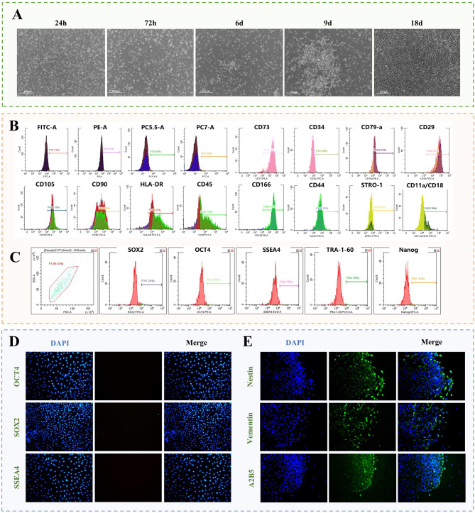
The isolated BSCs expressed mesenchymal stem cell-positive markers, including CD105, CD73, CD29, CD166, CD44, and CD90. Meanwhile, the mesenchymal stem cell-negative markers, including HLA-DR, CD45, and CD79a, were also found in BSCs. The BSCs did not express pluripotent stem cell markers, including SOX2, Nanog, OCT4, SSEA4, and TRA-1-60. Immunofluorescence detection showed that BSCs expressed neural stem/progenitor cell markers, including Vimentin, Nestin, and A2B5. Following BSC treatment, pathological improvements were observed in WMI. The expressions of mature OLs markers myelin basic protein and myelin-associated glycoprotein were increased in the corpus callosum and periventricular areas. Meanwhile, the numbers of myelin sheath increased, and learning and memory abilities improved. Furthermore, a decrease in B7-2+/Iba1 + proinflammatory microglia and an increase in CD206+/Iba1 + anti-inflammatory microglia were observed. The mRNA expressions of proinflammatory factors (Il1b, Il6, Ifng, and Tnfa) and anti-inflammatory factors (Arg1 and Tgfb) decreased and increased, respectively.
Our findings suggest that BSCs can improve the maturation of OLs following WMI in newborn rats. The mechanisms may be attributed to the reduced proinflammatory microglia cells and factors as well as the increased anti-inflammatory microglia cells and factors.
据报道,母乳干细胞(BSCs)对婴儿具有潜在益处。然而,BSCs是否能改善脑损伤尚不清楚。本研究建立了BSCs培养体系,并研究了BSCs在治疗白质损伤(WMI)中的作用。
收集分娩后1至5天健康哺乳期妇女的母乳样本。将BSCs在专用培养基中培养,然后通过流式细胞术和免疫荧光方法进行鉴定。通过结扎出生后第3天(P3)的Sprague-Dawley大鼠的右颈动脉并使其暴露于6%低氧环境2小时,建立WMI大鼠模型。大鼠分为假手术组、WMI+母乳细胞组(WMI+BC)和WMI+生理盐水组(WMI+NS)。在WMI+BC组中,建模后24小时将5μL BSCs(1×10)注入侧脑室。观察少突胶质细胞(OL)标志物的四个不同阶段。使用Morris水迷宫试验进行长期神经行为评估。检测炎性细胞因子和促炎性小胶质细胞的比例,以研究BSC治疗的机制。
分离出的BSCs表达间充质干细胞阳性标志物,包括CD105、CD73、CD29、CD166、CD44和CD90。同时,在BSCs中也发现了间充质干细胞阴性标志物,包括HLA-DR、CD45和CD79a。BSCs不表达多能干细胞标志物,包括SOX2、Nanog、OCT4、SSEA4和TRA-1-60。免疫荧光检测显示,BSCs表达神经干细胞/祖细胞标志物,包括波形蛋白、巢蛋白和A2B5。BSC治疗后,WMI出现病理改善。胼胝体和脑室周围区域成熟OL标志物髓鞘碱性蛋白和髓鞘相关糖蛋白的表达增加。同时,髓鞘数量增加,学习和记忆能力提高。此外,观察到B7-2+/Iba1+促炎性小胶质细胞减少,CD206+/Iba1+抗炎性小胶质细胞增加。促炎因子(Il1b、Il6、Ifng和Tnfa)和抗炎因子(Arg1和Tgfb)的mRNA表达分别降低和升高。
我们的研究结果表明,BSCs可以改善新生大鼠WMI后OL的成熟。其机制可能归因于促炎性小胶质细胞和因子减少以及抗炎性小胶质细胞和因子增加。